Sequence-structure mapping errors in the PDB: OB-fold domains
Venclovas, C., Ginalski, K., Kang, C.(2004) Protein Sci 13: 1594-1602
- PubMed: 15133161
- DOI: https://doi.org/10.1110/ps.04634604
- Primary Citation of Related Structures:
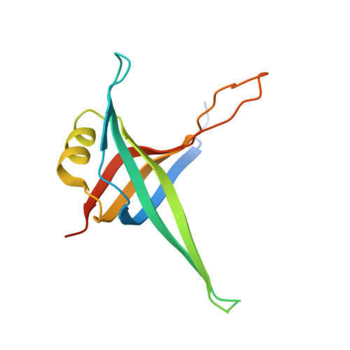
1S3O - PubMed Abstract:
The Protein Data Bank (PDB) is the single most important repository of structural data for proteins and other biologically relevant molecules. Therefore, it is critically important to keep the PDB data, as much as possible, error-free. In this study, we have analyzed PDB crystal structures possessing oligonucleotide/oligosaccharide binding (OB)-fold, one of the highly populated folds, for the presence of sequence-structure mapping errors. Using energy-based structure quality assessment coupled with sequence analyses, we have found that there are at least five OB-structures in the PDB that have regions where sequences have been incorrectly mapped onto the structure. We have demonstrated that the combination of these computation techniques is effective not only in detecting sequence-structure mapping errors, but also in providing guidance to correct them. Namely, we have used results of computational analysis to direct a revision of X-ray data for one of the PDB entries containing a fairly inconspicuous sequence-structure mapping error. The revised structure has been deposited with the PDB. We suggest use of computational energy assessment and sequence analysis techniques to facilitate structure determination when homologs having known structure are available to use as a reference. Such computational analysis may be useful in either guiding the sequence-structure assignment process or verifying the sequence mapping within poorly defined regions.
Organizational Affiliation:
Biology and Biotechnology Research Program, Lawrence Livermore National Laboratory, L-448, PO Box 808, Livermore, CA 94551, USA. venclovas@Ilnl.gov