1RDF

G50P mutant of phosphonoacetaldehyde hydrolase in complex with substrate analogue vinyl sulfonate


Experimental Data Snapshot

  • Method: X-RAY DIFFRACTION
  • Resolution: 2.80 Å
  • R-Value Free: 0.260 
  • R-Value Work: 0.230 
  • R-Value Observed: 0.230 

wwPDB Validation   3D Report Full Report


This is version 1.4 of the entry. See complete history


Literature

Analysis of the substrate specificity loop of the HAD superfamily cap domain

Lahiri, S.D.Zhang, G.Dai, J.Dunaway-Mariano, D.Allen, K.N.

(2004) Biochemistry 43: 2812-2820

  • DOI: https://doi.org/10.1021/bi0356810
  • Primary Citation of Related Structures:  
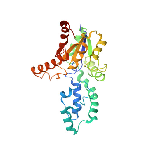
    1RDF

  • PubMed Abstract: 

    The haloacid dehalogenase (HAD) superfamily includes a variety of enzymes that catalyze the cleavage of substrate C-Cl, P-C, and P-OP bonds via nucleophilic substitution pathways. All members possess the alpha/beta core domain, and many also possess a small cap domain. The active site of the core domain is formed by four loops (corresponding to sequence motifs 1-4), which position substrate and cofactor-binding residues as well as the catalytic groups that mediate the "core" chemistry. The cap domain is responsible for the diversification of chemistry within the family. A tight beta-turn in the helix-loop-helix motif of the cap domain contains a stringently conserved Gly (within sequence motif 5), flanked by residues whose side chains contribute to the catalytic site formed at the domain-domain interface. To define the role of the conserved Gly in the structure and function of the cap domain loop of the HAD superfamily members phosphonoacetaldehyde hydrolase and beta-phosphoglucomutase, the Gly was mutated to Pro, Val, or Ala. The catalytic activity was severely reduced in each mutant. To examine the impact of Gly substitution on loop 5 conformation, the X-ray crystal structure of the Gly50Pro phosphonoacetaldehyde hydrolase mutant was determined. The altered backbone conformation at position 50 had a dramatic effect on the spatial disposition of the side chains of neighboring residues. Lys53, the Schiff Base forming lysine, had rotated out of the catalytic site and the side chain of Leu52 had moved to fill its place. On the basis of these studies, it was concluded that the flexibility afforded by the conserved Gly is critical to the function of loop 5 and that it is a marker by which the cap domain substrate specificity loop can be identified within the amino acid sequence of HAD family members.


  • Organizational Affiliation

    Department of Physiology and Biophysics, Boston University School of Medicine, Boston, Massachusetts 02118-2394, USA.


Macromolecules
Find similar proteins by:  (by identity cutoff)  |  3D Structure
Entity ID: 1
MoleculeChains Sequence LengthOrganismDetailsImage
phosphonoacetaldehyde hydrolase
A, B, C, D, E
A, B, C, D, E, F
267Bacillus cereusMutation(s): 1 
UniProt
Find proteins for O31156 (Bacillus cereus)
Explore O31156 
Go to UniProtKB:  O31156
Entity Groups  
Sequence Clusters30% Identity50% Identity70% Identity90% Identity95% Identity100% Identity
UniProt GroupO31156
Sequence Annotations
Expand
  • Reference Sequence
Small Molecules
Ligands 2 Unique
IDChains Name / Formula / InChI Key2D Diagram3D Interactions
ESA
Query on ESA

Download Ideal Coordinates CCD File 
H [auth A]
J [auth B]
L [auth C]
N [auth D]
P [auth E]
H [auth A],
J [auth B],
L [auth C],
N [auth D],
P [auth E],
R [auth F]
ETHANESULFONIC ACID
C2 H6 O3 S
CCIVGXIOQKPBKL-UHFFFAOYSA-N
MG
Query on MG

Download Ideal Coordinates CCD File 
G [auth A]
I [auth B]
K [auth C]
M [auth D]
O [auth E]
G [auth A],
I [auth B],
K [auth C],
M [auth D],
O [auth E],
Q [auth F]
MAGNESIUM ION
Mg
JLVVSXFLKOJNIY-UHFFFAOYSA-N
Experimental Data & Validation

Experimental Data

  • Method: X-RAY DIFFRACTION
  • Resolution: 2.80 Å
  • R-Value Free: 0.260 
  • R-Value Work: 0.230 
  • R-Value Observed: 0.230 
  • Space Group: C 1 2 1
Unit Cell:
Length ( Å )Angle ( ˚ )
a = 181.714α = 90
b = 147.348β = 125.21
c = 131.308γ = 90
Software Package:
Software NamePurpose
CNSrefinement
DENZOdata reduction
SCALEPACKdata scaling
AMoREphasing

Structure Validation

View Full Validation Report


Entry History 

Deposition Data

Revision History  (Full details and data files)

  • Version 1.0: 2004-08-31
    Type: Initial release
  • Version 1.1: 2008-04-29
    Changes: Version format compliance
  • Version 1.2: 2011-07-13
    Changes: Version format compliance
  • Version 1.3: 2021-10-27
    Changes: Database references, Derived calculations
  • Version 1.4: 2023-08-23
    Changes: Data collection, Refinement description