Crystal structure of Escherichia coli lytic transglycosylase Slt35 reveals a lysozyme-like catalytic domain with an EF-hand.
van Asselt, E.J., Dijkstra, A.J., Kalk, K.H., Takacs, B., Keck, W., Dijkstra, B.W.(1999) Structure 7: 1167-1180
- PubMed: 10545329
- DOI: https://doi.org/10.1016/s0969-2126(00)80051-9
- Primary Citation of Related Structures:
1QUS, 1QUT - PubMed Abstract:
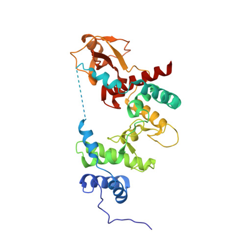
Lytic transglycosylases are bacterial muramidases that catalyse the cleavage of the beta- 1,4-glycosidic bond between N-acetylmuramic acid (MurNAc) and N-acetylglucosamine (GlcNAc) in peptidoglycan with concomitant formation of a 1,6-anhydrobond in the MurNAc residue. These muramidases play an important role in the metabolism of the bacterial cell wall and might therefore be potential targets for the rational design of antibacterial drugs. One of the lytic transglycosylases is Slt35, a naturally occurring soluble fragment of the outer membrane bound lytic transglycosylase B (MltB) from Escherichia coli. The crystal structure of Slt35 has been determined at 1.7 A resolution. The structure reveals an ellipsoid molecule with three domains called the alpha, beta and core domains. The core domain is sandwiched between the alpha and beta domains. Its fold resembles that of lysozyme, but it contains a single metal ion binding site in a helix-loop-helix module that is surprisingly similar to the eukaryotic EF-hand calcium-binding fold. Interestingly, the Slt35 EF-hand loop consists of 15 residues instead of the usual 12 residues. The only other prokaryotic proteins with an EF-hand motif identified so far are the D-galactose-binding proteins. Residues from the alpha and core domains form a deep groove where the substrate fragment GlcNAc can be bound. The three-domain structure of Slt35 is completely different from the Slt70 structure, the only other lytic transglycosylase of known structure. Nevertheless, the core domain of Slt35 closely resembles the fold of the catalytic domain of Slt70, despite the absence of any obvious sequence similarity. Residue Glu162 of Slt35 is in an equivalent position to Glu478, the catalytic acid/base of Slt70. GlcNAc binds close to Glu162 in the deep groove. Moreover, mutation of Glu162 into a glutamine residue yielded a completely inactive enzyme. These observations indicate the location of the active site and strongly support a catalytic role for Glu162.
Organizational Affiliation:
BIOSON Research Institute, Laboratory of Biophysical Chemistry Groningen University, Nijenborgh 4, 9747 AG, Groningen, The Netherlands.