1QTN

CRYSTAL STRUCTURE OF THE COMPLEX OF CASPASE-8 WITH THE TETRAPEPTIDE INHIBITOR ACE-IETD-ALDEHYDE


Experimental Data Snapshot

  • Method: X-RAY DIFFRACTION
  • Resolution: 1.20 Å
  • R-Value Free: 0.188 
  • R-Value Work: 0.143 
  • R-Value Observed: 0.166 

wwPDB Validation   3D Report Full Report


This is version 1.6 of the entry. See complete history


Literature

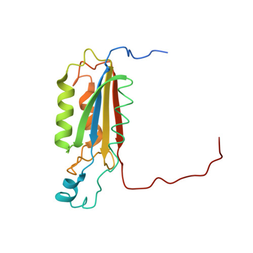
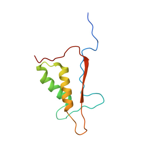
The atomic-resolution structure of human caspase-8, a key activator of apoptosis.

Watt, W.Koeplinger, K.A.Mildner, A.M.Heinrikson, R.L.Tomasselli, A.G.Watenpaugh, K.D.

(1999) Structure 7: 1135-1143

  • DOI: https://doi.org/10.1016/s0969-2126(99)80180-4
  • Primary Citation of Related Structures:  
    1QTN

  • PubMed Abstract: 

    Caspases are a family of cysteine proteases that have important intracellular roles in inflammation and apoptosis. Caspase-8 activates downstream caspases which are unable to carry out autocatalytic processing and activation. Caspase-8 is designated as an initiator caspase and is believed to sit at the apex of the Fas- or TNF-mediated apoptotic cascade. In view of this role, the enzyme is an attractive target for the design of inhibitors aimed at blocking the undesirable cell death associated with a range of degenerative disorders. The structure of recombinant human caspase-8, covalently modified with the inhibitor acetyl-Ile-Glu-Thr-Asp-aldehyde, has been determined by X-ray crystallography to 1.2 A resolution. The asymmetric unit contains the p18-p11 heterodimer; the biologically important molecule contains two dimers. The overall fold is very similar to that of caspase-1 and caspase-3, but significant differences exist in the substrate-binding region. The structure answers questions about the enzyme-inhibitor complex that could not be explained from earlier caspase structures solved at lower resolution. The catalytic triad in caspase-8 comprises Cys360, His317 and the backbone carbonyl oxygen atom of Arg258, which points towards the Nepsilon atom of His317. The oxygen atom attached to the tetrahedral carbon in the thiohemiacetal group of the inhibitor is hydrogen bonded to Ndelta of His317, and is not in a region characteristic of a classical 'oxyanion hole'. The N-acetyl group of the inhibitor is in the trans configuration. The caspase-8-inhibitor structure provides the basis for understanding structure/function relationships in this important initiator of the proteolytic cascade that leads to programmed cell death.


  • Organizational Affiliation

    Structural, Analytical & Medicinal Chemistry Pharmacia & Upjohn Inc. 301 Henrietta Street, Kalamazoo, MI 49007, USA.


Macromolecules
Find similar proteins by:  (by identity cutoff)  |  3D Structure
Entity ID: 1
MoleculeChains Sequence LengthOrganismDetailsImage
CASPASE-8164Homo sapiensMutation(s): 0 
EC: 3.4.22
UniProt & NIH Common Fund Data Resources
Find proteins for Q14790 (Homo sapiens)
Explore Q14790 
Go to UniProtKB:  Q14790
PHAROS:  Q14790
GTEx:  ENSG00000064012 
Entity Groups  
Sequence Clusters30% Identity50% Identity70% Identity90% Identity95% Identity100% Identity
UniProt GroupQ14790
Sequence Annotations
Expand
  • Reference Sequence
Find similar proteins by:  (by identity cutoff)  |  3D Structure
Entity ID: 2
MoleculeChains Sequence LengthOrganismDetailsImage
CASPASE-895Homo sapiensMutation(s): 0 
EC: 3.4.22
UniProt & NIH Common Fund Data Resources
Find proteins for Q14790 (Homo sapiens)
Explore Q14790 
Go to UniProtKB:  Q14790
PHAROS:  Q14790
GTEx:  ENSG00000064012 
Entity Groups  
Sequence Clusters30% Identity50% Identity70% Identity90% Identity95% Identity100% Identity
UniProt GroupQ14790
Sequence Annotations
Expand
  • Reference Sequence

Find similar proteins by:  Sequence   |   3D Structure  

Entity ID: 3
MoleculeChains Sequence LengthOrganismDetailsImage
ACETYL-ILE-GLU-THR-ASP-ALDEHYDE5N/AMutation(s): 0 
Sequence Annotations
Expand
  • Reference Sequence
Small Molecules
Ligands 1 Unique
IDChains Name / Formula / InChI Key2D Diagram3D Interactions
DTD
Query on DTD

Download Ideal Coordinates CCD File 
D [auth A],
E [auth B]
DITHIANE DIOL
C4 H8 O2 S2
YPGMOWHXEQDBBV-IMJSIDKUSA-N
Biologically Interesting Molecules (External Reference) 1 Unique
Experimental Data & Validation

Experimental Data

  • Method: X-RAY DIFFRACTION
  • Resolution: 1.20 Å
  • R-Value Free: 0.188 
  • R-Value Work: 0.143 
  • R-Value Observed: 0.166 
  • Space Group: P 31 2 1
Unit Cell:
Length ( Å )Angle ( ˚ )
a = 62.4α = 90
b = 62.4β = 90
c = 129.25γ = 120
Software Package:
Software NamePurpose
AMoREphasing
SHELXL-97refinement
SMARTdata reduction
SAINTdata scaling

Structure Validation

View Full Validation Report


Entry History 

Deposition Data

Revision History  (Full details and data files)

  • Version 1.0: 2000-09-20
    Type: Initial release
  • Version 1.1: 2008-04-27
    Changes: Version format compliance
  • Version 1.2: 2011-07-13
    Changes: Atomic model, Database references, Derived calculations, Non-polymer description, Structure summary, Version format compliance
  • Version 1.3: 2012-04-04
    Changes: Structure summary
  • Version 1.4: 2012-12-12
    Changes: Other
  • Version 1.5: 2023-08-16
    Changes: Data collection, Database references, Derived calculations, Refinement description
  • Version 1.6: 2023-11-15
    Changes: Data collection