1QC1

CRYSTAL STRUCTURE OF THE SELF-FITTED B-DNA DECAMER D(CCGCCGGCGG)

  • Classification: DNA
  • Mutation(s): No 

  • Deposited: 1999-05-14 Released: 1999-05-28 
  • Deposition Author(s): Timsit, Y., Moras, D.

Experimental Data Snapshot

  • Method: X-RAY DIFFRACTION
  • Resolution: 2.50 Å
  • R-Value Observed: 0.147 

wwPDB Validation   3D Report Full Report


This is version 1.4 of the entry. See complete history


Literature

DNA self-fitting: the double helix directs the geometry of its supramolecular assembly

Timsit, Y.Moras, D.

(1994) EMBO J 13: 2737-2746

  • DOI: https://doi.org/10.1002/j.1460-2075.1994.tb06567.x
  • Primary Citation of Related Structures:  
    1QC1

  • PubMed Abstract: 

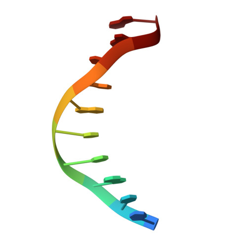
    Groove-backbone interaction is a natural and biologically relevant mechanism for the specific assembly of B-DNA double helices. Crystal engineering and crystal packing analysis of oligonucleotides of different sizes and sequences reveal that the sequence-dependent self-fitting of B-DNA helices is a dominant constraint for their ordered assembly. It can override the other intermolecular interactions and impose the overall geometry of the packing. Analysis of experimental examples of architectural motifs formed by the geometric combination of self-fitted DNA segments leads to general rules for DNA assembly. Like a directing piece for a supramolecular 'construction set', the double helix imposes a limited number of geometric solutions. These basic architectural constraints could direct, in a codified manner, the formation of higher-order structures. DNA architectural motifs exhibit new structural and electrostatic properties which could have some implications for their molecular recognition by proteins acting on DNA.


  • Organizational Affiliation

    UPR de Biologie Structurale, CNRS, Institut de Biologie Moléculaire et Cellulaire, Strasbourg, France.


Macromolecules

Find similar nucleic acids by:  Sequence   |   3D Structure  

Entity ID: 1
MoleculeChains LengthOrganismImage
5'-D(*CP*CP*GP*CP*CP*GP*GP*CP*GP*G)-3'
A, B
10N/A
Sequence Annotations
Expand
  • Reference Sequence
Experimental Data & Validation

Experimental Data

  • Method: X-RAY DIFFRACTION
  • Resolution: 2.50 Å
  • R-Value Observed: 0.147 
  • Space Group: H 3
Unit Cell:
Length ( Å )Angle ( ˚ )
a = 53.7α = 90
b = 53.7β = 90
c = 45γ = 120
Software Package:
Software NamePurpose
X-GENdata scaling
X-GENdata reduction
NUCLSQrefinement

Structure Validation

View Full Validation Report


Entry History 

Deposition Data

Revision History  (Full details and data files)

  • Version 1.0: 1999-05-28
    Type: Initial release
  • Version 1.1: 2008-04-27
    Changes: Version format compliance
  • Version 1.2: 2011-07-13
    Changes: Version format compliance
  • Version 1.3: 2017-10-04
    Changes: Refinement description
  • Version 1.4: 2024-02-14
    Changes: Data collection, Database references, Derived calculations