1PUE

PU.1 ETS DOMAIN-DNA COMPLEX


Experimental Data Snapshot

  • Method: X-RAY DIFFRACTION
  • Resolution: 2.10 Å
  • R-Value Free: 0.287 
  • R-Value Work: 0.225 
  • R-Value Observed: 0.225 

wwPDB Validation   3D Report Full Report


This is version 1.4 of the entry. See complete history


Literature

A new pattern for helix-turn-helix recognition revealed by the PU.1 ETS-domain-DNA complex.

Kodandapani, R.Pio, F.Ni, C.Z.Piccialli, G.Klemsz, M.McKercher, S.Maki, R.A.Ely, K.R.

(1996) Nature 380: 456-460

  • DOI: https://doi.org/10.1038/380456a0
  • Primary Citation of Related Structures:  
    1PUE

  • PubMed Abstract: 

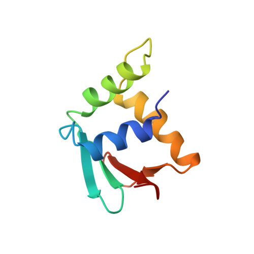
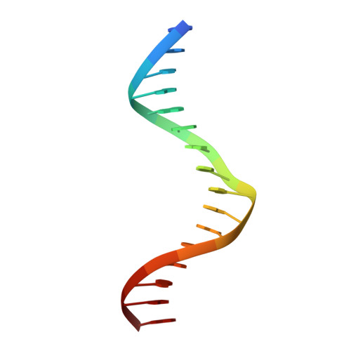
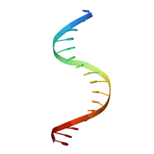
    The Ets family of transcription factors, of which there are now about 35 members regulate gene expression during growth and development. They share a conserved domain of around 85 amino acids which binds as a monomer to the DNA sequence 5'-C/AGGAA/T-3'. We have determined the crystal structure of an ETS domain complexed with DNA, at 2.3-A resolution. The domain is similar to alpha + beta (winged) 'helix-turn-helix' proteins and interacts with a ten-base-pair region of duplex DNA which takes up a uniform curve of 8 degrees. The domain contacts the DNA by a novel loop-helix-loop architecture. Four of amino acids that directly interact with the DNA are highly conserved: two arginines from the recognition helix lying in the major groove, one lysine from the 'wing' that binds upstream of the core GGAA sequence, and another lysine, from the 'turn' of the 'helix-turn-helix' motif, which binds downstream and on the opposite strand.


  • Organizational Affiliation

    La Jolla Cancer Research Center at the Burnham Institute, California 92037, USA.


Macromolecules

Find similar proteins by:  (by identity cutoff)  |  3D Structure
Entity ID: 3
MoleculeChains Sequence LengthOrganismDetailsImage
PROTEIN (TRANSCRIPTION FACTOR PU.1 (TF PU.1))
E, F
89Mus musculusMutation(s): 0 
UniProt & NIH Common Fund Data Resources
Find proteins for P17433 (Mus musculus)
Explore P17433 
Go to UniProtKB:  P17433
IMPC:  MGI:98282
Entity Groups  
Sequence Clusters30% Identity50% Identity70% Identity90% Identity95% Identity100% Identity
UniProt GroupP17433
Sequence Annotations
Expand
  • Reference Sequence

Find similar nucleic acids by:  Sequence   |   3D Structure  

Entity ID: 1
MoleculeChains LengthOrganismImage
DNA (5'-D(*AP*AP*AP*AP*AP*GP*GP*GP*GP*AP*AP*GP*TP*GP*GP*G)-3')
A, C
16synthetic construct
Sequence Annotations
Expand
  • Reference Sequence

Find similar nucleic acids by:  Sequence   |   3D Structure  

Entity ID: 2
MoleculeChains LengthOrganismImage
DNA (5'-D(*TP*CP*CP*CP*AP*CP*TP*TP*CP*CP*CP*CP*TP*TP*TP*T)-3')
B, D
16synthetic construct
Sequence Annotations
Expand
  • Reference Sequence
Experimental Data & Validation

Experimental Data

  • Method: X-RAY DIFFRACTION
  • Resolution: 2.10 Å
  • R-Value Free: 0.287 
  • R-Value Work: 0.225 
  • R-Value Observed: 0.225 
  • Space Group: C 1 2 1
Unit Cell:
Length ( Å )Angle ( ˚ )
a = 89.1α = 90
b = 101.9β = 111.2
c = 55.6γ = 90
Software Package:
Software NamePurpose
X-PLORrefinement
MOSFLMdata reduction

Structure Validation

View Full Validation Report


Entry History 

Deposition Data

Revision History  (Full details and data files)

  • Version 1.0: 1997-02-12
    Type: Initial release
  • Version 1.1: 2008-05-22
    Changes: Version format compliance
  • Version 1.2: 2011-07-13
    Changes: Version format compliance
  • Version 1.3: 2017-02-15
    Changes: Source and taxonomy, Structure summary
  • Version 1.4: 2024-02-14
    Changes: Data collection, Database references