1PL3

Cytochrome Domain Of Cellobiose Dehydrogenase, M65H mutant


Experimental Data Snapshot

  • Method: X-RAY DIFFRACTION
  • Resolution: 1.90 Å
  • R-Value Free: 0.198 
  • R-Value Work: 0.173 
  • R-Value Observed: 0.174 

wwPDB Validation   3D Report Full Report


Ligand Structure Quality Assessment 


This is version 3.2 of the entry. See complete history


Literature

Biophysical and Structural Analysis of a Novel Heme b Iron Ligation in the Flavocytochrome Cellobiose Dehydrogenase.

Rotsaert, F.A.J.Hallberg, B.M.De Vries, S.Moenne-Loccoz, P.Divne, C.Renganathan, V.Gold, M.H.

(2003) J Biol Chem 278: 33224-33231

  • DOI: https://doi.org/10.1074/jbc.M302653200
  • Primary Citation of Related Structures:  
    1PL3

  • PubMed Abstract: 

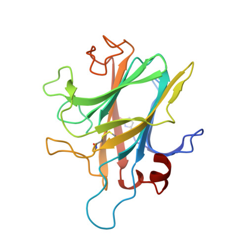
    The fungal extracellular flavocytochrome cellobiose dehydrogenase (CDH) participates in lignocellulose degradation. The enzyme has a cytochrome domain connected to a flavin-binding domain by a peptide linker. The cytochrome domain contains a 6-coordinate low spin b-type heme with unusual iron ligands and coordination geometry. Wild type CDH is only the second example of a b-type heme with Met-His ligation, and it is the first example of a Met-His ligation of heme b where the ligands are arranged in a nearly perpendicular orientation. To investigate the ligation further, Met65 was replaced with a histidine to create a bis-histidyl ligated iron typical of b-type cytochromes. The variant is expressed as a stable 90-kDa protein that retains the flavin domain catalytic reactivity. However, the ability of the mutant to reduce external one-electron acceptors such as cytochrome c is impaired. Electrochemical measurements demonstrate a decrease in the redox midpoint potential of the heme by 210 mV. In contrast to the wild type enzyme, the ferric state of the protoheme displays a mixed low spin/high spin state at room temperature and low spin character at 90 K, as determined by resonance Raman spectroscopy. The wild type cytochrome does not bind CO, but the ferrous state of the variant forms a CO complex, although the association rate is very low. The crystal structure of the M65H cytochrome domain has been determined at 1.9 A resolution. The variant structure confirms a bis-histidyl ligation but reveals unusual features. As for the wild type enzyme, the ligands have a nearly perpendicular arrangement. Furthermore, the iron is bound by imidazole N delta 1 and N epsilon 2 nitrogen atoms, rather than the typical N epsilon 2/N epsilon 2 coordination encountered in bis-histidyl ligated heme proteins. To our knowledge, this is the first example of a bis-histidyl N delta 1/N epsilon 2-coordinated protoporphyrin IX iron.


  • Organizational Affiliation

    Department of Biochemistry and Molecular Biology, OGI School of Science and Engineering at Oregon Health & Science University, Beaverton, Oregon 97006-8921, USA.


Macromolecules
Find similar proteins by:  (by identity cutoff)  |  3D Structure
Entity ID: 1
MoleculeChains Sequence LengthOrganismDetailsImage
Cellobiose dehydrogenase
A, B
186Phanerodontia chrysosporiumMutation(s): 1 
Gene Names: CDH-1 AND CDH-2
EC: 1.1.99.18
UniProt
Find proteins for Q01738 (Phanerodontia chrysosporium)
Explore Q01738 
Go to UniProtKB:  Q01738
Entity Groups  
Sequence Clusters30% Identity50% Identity70% Identity90% Identity95% Identity100% Identity
UniProt GroupQ01738
Sequence Annotations
Expand
  • Reference Sequence
Oligosaccharides

Help

Entity ID: 2
MoleculeChains Length2D Diagram Glycosylation3D Interactions
2-acetamido-2-deoxy-beta-D-glucopyranose-(1-4)-2-acetamido-2-deoxy-beta-D-glucopyranose
C, D
2N-Glycosylation
Glycosylation Resources
GlyTouCan:  G42666HT
GlyCosmos:  G42666HT
GlyGen:  G42666HT
Small Molecules
Ligands 3 Unique
IDChains Name / Formula / InChI Key2D Diagram3D Interactions
HEM
Query on HEM

Download Ideal Coordinates CCD File 
I [auth A],
M [auth B]
PROTOPORPHYRIN IX CONTAINING FE
C34 H32 Fe N4 O4
KABFMIBPWCXCRK-RGGAHWMASA-L
1PG
Query on 1PG

Download Ideal Coordinates CCD File 
J [auth A]2-(2-{2-[2-(2-METHOXY-ETHOXY)-ETHOXY]-ETHOXY}-ETHOXY)-ETHANOL
C11 H24 O6
SLNYBUIEAMRFSZ-UHFFFAOYSA-N
CD
Query on CD

Download Ideal Coordinates CCD File 
E [auth A]
F [auth A]
G [auth A]
H [auth A]
K [auth B]
E [auth A],
F [auth A],
G [auth A],
H [auth A],
K [auth B],
L [auth B]
CADMIUM ION
Cd
WLZRMCYVCSSEQC-UHFFFAOYSA-N
Modified Residues  1 Unique
IDChains TypeFormula2D DiagramParent
PCA
Query on PCA
A, B
L-PEPTIDE LINKINGC5 H7 N O3GLN
Experimental Data & Validation

Experimental Data

  • Method: X-RAY DIFFRACTION
  • Resolution: 1.90 Å
  • R-Value Free: 0.198 
  • R-Value Work: 0.173 
  • R-Value Observed: 0.174 
  • Space Group: P 65
Unit Cell:
Length ( Å )Angle ( ˚ )
a = 139.033α = 90
b = 139.033β = 90
c = 52.668γ = 120
Software Package:
Software NamePurpose
REFMACrefinement
SCALAdata scaling
CNSrefinement
MOSFLMdata reduction
CCP4data scaling
CNSphasing

Structure Validation

View Full Validation Report


Ligand Structure Quality Assessment 


Entry History 

Deposition Data

Revision History  (Full details and data files)

  • Version 1.0: 2003-07-01
    Type: Initial release
  • Version 1.1: 2008-04-29
    Changes: Version format compliance
  • Version 1.2: 2011-07-13
    Changes: Non-polymer description, Version format compliance
  • Version 1.3: 2019-07-24
    Changes: Data collection, Derived calculations, Refinement description
  • Version 2.0: 2019-12-25
    Changes: Database references, Derived calculations, Polymer sequence
  • Version 3.0: 2020-07-29
    Type: Remediation
    Reason: Carbohydrate remediation
    Changes: Atomic model, Data collection, Derived calculations, Structure summary
  • Version 3.1: 2021-10-27
    Changes: Database references, Structure summary
  • Version 3.2: 2023-08-16
    Changes: Data collection, Refinement description