Solution Structure of the Esperamicin A1-DNA Complex
Kumar, R.A., Ikemoto, N., Patel, D.J.(1997) J Mol Biol 265: 173-186
- PubMed: 9020981
- DOI: https://doi.org/10.1006/jmbi.1996.0719
- Primary Citation of Related Structures:
1PIK - PubMed Abstract:
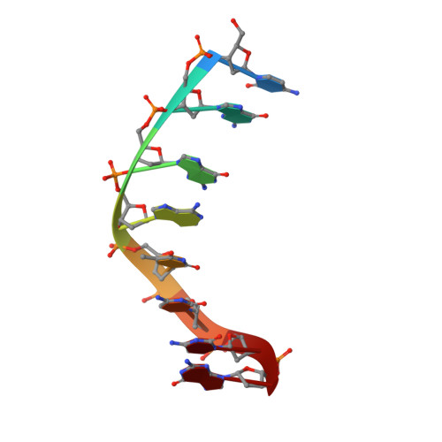
Esperamicin A1 is an enediyne antibiotic possessing antitumor activity associated with its ability to bind and, following activation, affect strand cleavage of DNA. We report on the solution structure of the esperamicin A1-d(C-G-G-A-T-C-C-G) duplex complex based on a combined analysis of NMR and molecular dynamics calculations including intensity refinement in a water box. The refined solution structures of the complex provide a molecular explanation of the sequence specificity for binding and cleavage by this member of the enediyne family of antitumor antibiotics. Esperamicin A1 binds to the DNA minor groove with its methoxyacrylyl-anthranilate moiety intercalating into the helix at the (G2-G3)-(C6'-C7') step. The methoxyacrylyl-anthranilate intercalator and the minor groove binding A-B-C+ risaccharide moieties rigidly anchor the enediyne in the minor groove such that the pro-radical centers of the enediyne are proximal to their anticipated proton abstraction sites. Specifically, the pro-radical C-3 and C-6 atoms are aligned opposite the abstractable H-5' (pro-S) proton of C6 and the H-1' proton of C6' on partner strands, respectively, in the complex. The thiomethyl sugar B residue is buried deep in an edgewise manner in the minor groove with its two faces sandwiched between the walls of the groove. Further, the polarizable sulfur atom of the thiomethyl group of sugar B residue is positioned opposite and can hydrogen-bond to the exposed amino proton of G3' in the complex. There is little perturbation away from a right-handed Watson-Crick base-paired duplex in the complex other than unwinding of the helix at the intercalation site and widening of the minor groove centered about the enediyne-binding and anthranilate intercalation sites. Sequence-specific binding of esperamicin A1 to the d(C-G-G-A-T-C-C-G) duplex is favored by the complementarity of the fit between the drug and the floor of the minor groove, good stacking between the intercalating anthranilate ring and flanking purine bases and intermolecular hydrogen-bonding interactions.
Organizational Affiliation:
Cellular Biochemistry and Biophysics Program Memorial Sloan-Kettering Cancer Center, New York, NY 10021, USA.