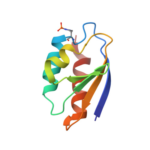
High-resolution structure of the phosphorylated form of the histidine-containing phosphocarrier protein HPr from Escherichia coli determined by restrained molecular dynamics from NMR-NOE data.
van Nuland, N.A., Boelens, R., Scheek, R.M., Robillard, G.T.(1995) J Mol Biol 246: 180-193
- PubMed: 7853396
- DOI: https://doi.org/10.1006/jmbi.1994.0075
- Primary Citation of Related Structures:
1PFH - PubMed Abstract:
The solution structure of the phosphorylated form of the histidine-containing phosphocarrier protein, HPr, from Escherichia coli has been determined by NMR in combination with restrained molecular dynamics simulations. The structure of phospho-HPr (P-HPr) results from a molecular dynamics simulation in water, using time-dependent distance restraints to attain agreement with the measured NOEs. Experimental restraints were identified from both three-dimensional 1H-1H-15N HSQC-NOESY and two-dimensional 1H-1HNOESY spectra, and compared with those of the unphosphorylated form. Structural changes upon phosphorylation of HPr are limited to the active site, as evidenced by changes in chemical shifts, in 3JNHH alpha-coupling constants and NOE patterns. Chemical shift changes were obtained mainly for protons that were positioned close to the phosphoryl group attached to the His15 imidazole ring. Differences could be detected in the intensity of the NOEs involving the side-chain protons of His15 and Pro18, resulting from a change in the relative position of the two rings. In addition, a small change could be detected in the three-bond J-coupling between the amide proton and the H alpha proton of Thr16 and Arg17 upon phosphorylation, in agreement with the changes of the phi torsion angle of these two residues obtained from time-averaged restrained molecular dynamics simulations in water. The proposed role of the torsion-angle strain at residue 16 in the mechanism of Streptococcus faecalis HPr is not supported by these results. In contrast, phosphorylation seems to introduce torsion angle strain at residue His15. This strain could facilitate the transfer of the phosphoryl group to the A-domain at enzyme II. The phospho-histidine is not stabilised by hydrogen bonds to the side-chain group of Arg17; instead stable hydrogen bonds are formed between the phosphate group and the backbone amide protons of Thr16 and Arg17, which show the largest changes in chemical shift upon phosphorylation, and a hydrogen bond involving the side-chain O gamma proton of Thr16. HPr accepts the phosphoryl group from enzyme I and donates it subsequently to the A domain of various enzyme II species. The binding site for EI on HPr resembles that of the A domain of the mannitol-specific enzyme II, as can be concluded from the changes on the amide proton and nitrogen chemical shifts observed via heteromolecular single-quantum coherence spectroscopy.
Organizational Affiliation:
Groningen Biomolecular Sciences and Biotechnology Institute, The Netherlands.