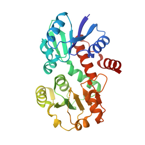
1P77

CRYSTAL STRUCTURE OF SHIKIMATE DEHYDROGENASE (AROE) FROM HAEMOPHILUS INFLUENZAE


Experimental Data Snapshot

  • Method: X-RAY DIFFRACTION
  • Resolution: 1.95 Å
  • R-Value Free: 0.244 
  • R-Value Work: 0.203 
  • R-Value Observed: 0.205 

wwPDB Validation   3D Report Full Report


This is version 2.0 of the entry. See complete history


Literature

The crystal structure of shikimate dehydrogenase (AroE) reveals a unique NADPH binding mode

Ye, S.von Delft, F.Brooun, A.Knuth, M.W.Swanson, R.V.McRee, D.E.

(2003) J Bacteriol 185: 4144-4151

  • DOI: https://doi.org/10.1128/JB.185.14.4144-4151.2003
  • Primary Citation of Related Structures:  
    1P74, 1P77

  • PubMed Abstract: 

    Shikimate dehydrogenase catalyzes the NADPH-dependent reversible reduction of 3-dehydroshikimate to shikimate. We report the first X-ray structure of shikimate dehydrogenase from Haemophilus influenzae to 2.4-A resolution and its complex with NADPH to 1.95-A resolution. The molecule contains two domains, a catalytic domain with a novel open twisted alpha/beta motif and an NADPH binding domain with a typical Rossmann fold. The enzyme contains a unique glycine-rich P-loop with a conserved sequence motif, GAGGXX, that results in NADPH adopting a nonstandard binding mode with the nicotinamide and ribose moieties disordered in the binary complex. A deep pocket with a narrow entrance between the two domains, containing strictly conserved residues primarily contributed by the catalytic domain, is identified as a potential 3-dehydroshikimate binding pocket. The flexibility of the nicotinamide mononucleotide portion of NADPH may be necessary for the substrate 3-dehydroshikimate to enter the pocket and for the release of the product shikimate.


  • Organizational Affiliation

    Syrrx Inc., San Diego, California 92121, USA.


Macromolecules
Find similar proteins by:  (by identity cutoff)  |  3D Structure
Entity ID: 1
MoleculeChains Sequence LengthOrganismDetailsImage
Shikimate 5-dehydrogenase272Haemophilus influenzaeMutation(s): 0 
Gene Names: AROE OR HI0655
EC: 1.1.1.25
UniProt
Find proteins for P43876 (Haemophilus influenzae (strain ATCC 51907 / DSM 11121 / KW20 / Rd))
Explore P43876 
Go to UniProtKB:  P43876
Entity Groups  
Sequence Clusters30% Identity50% Identity70% Identity90% Identity95% Identity100% Identity
UniProt GroupP43876
Sequence Annotations
Expand
  • Reference Sequence
Small Molecules
Ligands 2 Unique
IDChains Name / Formula / InChI Key2D Diagram3D Interactions
ATR
Query on ATR

Download Ideal Coordinates CCD File 
C [auth A]2'-MONOPHOSPHOADENOSINE-5'-DIPHOSPHATE
C10 H16 N5 O13 P3
YPTPYQSAVGGMFN-KQYNXXCUSA-N
ACT
Query on ACT

Download Ideal Coordinates CCD File 
B [auth A]ACETATE ION
C2 H3 O2
QTBSBXVTEAMEQO-UHFFFAOYSA-M
Experimental Data & Validation

Experimental Data

  • Method: X-RAY DIFFRACTION
  • Resolution: 1.95 Å
  • R-Value Free: 0.244 
  • R-Value Work: 0.203 
  • R-Value Observed: 0.205 
  • Space Group: C 2 2 21
Unit Cell:
Length ( Å )Angle ( ˚ )
a = 84.054α = 90
b = 82.728β = 90
c = 82.365γ = 90
Software Package:
Software NamePurpose
REFMACrefinement
HKL-2000data reduction
SCALEPACKdata scaling
AMoREphasing

Structure Validation

View Full Validation Report


Entry History 

Deposition Data

Revision History  (Full details and data files)

  • Version 1.0: 2003-08-12
    Type: Initial release
  • Version 1.1: 2008-04-29
    Changes: Version format compliance
  • Version 1.2: 2011-07-13
    Changes: Version format compliance
  • Version 1.3: 2018-01-31
    Changes: Database references
  • Version 2.0: 2023-08-16
    Changes: Atomic model, Data collection, Database references, Derived calculations, Refinement description