1P0Z

Sensor Kinase CitA binding domain


Experimental Data Snapshot

  • Method: X-RAY DIFFRACTION
  • Resolution: 1.60 Å
  • R-Value Free: 0.190 
  • R-Value Work: 0.168 
  • R-Value Observed: 0.168 

wwPDB Validation   3D Report Full Report


This is version 1.5 of the entry. See complete history


Literature

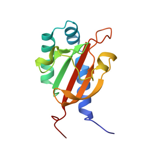
The structure of the periplasmic ligand-binding domain of the sensor kinase CitA reveals the first extracellular PAS domain.

Reinelt, S.Hofmann, E.Gerharz, T.Bott, M.Madden, D.R.

(2003) J Biol Chem 278: 39189-39196

  • DOI: https://doi.org/10.1074/jbc.M305864200
  • Primary Citation of Related Structures:  
    1P0Z

  • PubMed Abstract: 

    The integral membrane sensor kinase CitA of Klebsiella pneumoniae is part of a two-component signal transduction system that regulates the transport and metabolism of citrate in response to its environmental concentration. Two-component systems are widely used by bacteria for such adaptive processes, but the stereochemistry of periplasmic ligand binding and the mechanism of signal transduction across the membrane remain poorly understood. The crystal structure of the CitAP periplasmic sensor domain in complex with citrate reveals a PAS fold, a versatile ligand-binding structural motif that has not previously been observed outside the cytoplasm or implicated in the transduction of conformational signals across the membrane. Citrate is bound in a pocket that is shared among many PAS domains but that shows structural variation according to the nature of the bound ligand. In CitAP, some of the citrate contact residues are located in the final strand of the central beta-sheet, which is connected to the C-terminal transmembrane helix. These secondary structure elements thus provide a potential conformational link between the periplasmic ligand binding site and the cytoplasmic signaling domains of the receptor.


  • Organizational Affiliation

    Ion Channel Structure Group, Max Planck Institute for Medical Research, D-69120 Heidelberg, Germany.


Macromolecules
Find similar proteins by:  (by identity cutoff)  |  3D Structure
Entity ID: 1
MoleculeChains Sequence LengthOrganismDetailsImage
Sensor kinase citA
A, B, C, D, E
A, B, C, D, E, F, G, H, I, J
131Klebsiella pneumoniaeMutation(s): 0 
Gene Names: CITA
EC: 2.7.13.3
UniProt
Find proteins for P52687 (Klebsiella pneumoniae)
Explore P52687 
Go to UniProtKB:  P52687
Entity Groups  
Sequence Clusters30% Identity50% Identity70% Identity90% Identity95% Identity100% Identity
UniProt GroupP52687
Sequence Annotations
Expand
  • Reference Sequence
Small Molecules
Ligands 4 Unique
IDChains Name / Formula / InChI Key2D Diagram3D Interactions
MO7
Query on MO7

Download Ideal Coordinates CCD File 
CA [auth E]
GA [auth F]
KA [auth G]
M [auth A]
OA [auth H]
CA [auth E],
GA [auth F],
KA [auth G],
M [auth A],
OA [auth H],
Q [auth B],
SA [auth I],
U [auth C],
WA [auth J],
Y [auth D]
bis(mu4-oxo)-bis(mu3-oxo)-octakis(mu2-oxo)-dodecaoxo-heptamolybdenum (VI)
Mo7 O24
CFQDGBTUZUJXQE-UHFFFAOYSA-N
FLC
Query on FLC

Download Ideal Coordinates CCD File 
AA [auth E]
EA [auth F]
IA [auth G]
K [auth A]
MA [auth H]
AA [auth E],
EA [auth F],
IA [auth G],
K [auth A],
MA [auth H],
O [auth B],
QA [auth I],
S [auth C],
UA [auth J],
W [auth D]
CITRATE ANION
C6 H5 O7
KRKNYBCHXYNGOX-UHFFFAOYSA-K
OMO
Query on OMO

Download Ideal Coordinates CCD File 
DA [auth E]
HA [auth F]
LA [auth G]
N [auth A]
PA [auth H]
DA [auth E],
HA [auth F],
LA [auth G],
N [auth A],
PA [auth H],
R [auth B],
TA [auth I],
V [auth C],
XA [auth J],
Z [auth D]
MO(VI)(=O)(OH)2 CLUSTER
H2 Mo O3
GKDPEXRCAVYDOG-UHFFFAOYSA-L
NA
Query on NA

Download Ideal Coordinates CCD File 
BA [auth E]
FA [auth F]
JA [auth G]
L [auth A]
NA [auth H]
BA [auth E],
FA [auth F],
JA [auth G],
L [auth A],
NA [auth H],
P [auth B],
RA [auth I],
T [auth C],
VA [auth J],
X [auth D]
SODIUM ION
Na
FKNQFGJONOIPTF-UHFFFAOYSA-N
Experimental Data & Validation

Experimental Data

  • Method: X-RAY DIFFRACTION
  • Resolution: 1.60 Å
  • R-Value Free: 0.190 
  • R-Value Work: 0.168 
  • R-Value Observed: 0.168 
  • Space Group: P 1
Unit Cell:
Length ( Å )Angle ( ˚ )
a = 61.613α = 112.83
b = 84.333β = 107.36
c = 97.71γ = 93.74
Software Package:
Software NamePurpose
CNSrefinement
XDSdata reduction
XSCALEdata scaling
SHELXDphasing

Structure Validation

View Full Validation Report


Entry History 

Deposition Data

Revision History  (Full details and data files)

  • Version 1.0: 2003-08-19
    Type: Initial release
  • Version 1.1: 2008-04-01
    Changes: Version format compliance
  • Version 1.2: 2011-07-13
    Changes: Version format compliance
  • Version 1.3: 2014-04-09
    Changes: Non-polymer description
  • Version 1.4: 2014-09-10
    Changes: Database references
  • Version 1.5: 2024-02-14
    Changes: Data collection, Database references, Derived calculations