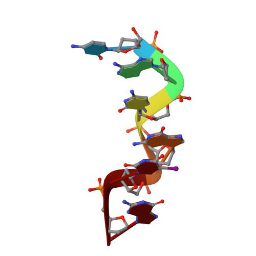
Exploration of the influence of 5-iodo-2'-deoxyuridine incorporation on the structure of d[CACG(IDU)G].
Schuerman, G., Van Hecke, K., Van Meervelt, L.(2003) Acta Crystallogr D Biol Crystallogr 59: 1525-1528
- PubMed: 12876373
- DOI: https://doi.org/10.1107/s0907444903012381
- Primary Citation of Related Structures:
1OMK - PubMed Abstract:
The first antiviral nucleoside 5-iodo-2'-deoxyuridine (IDU) against herpes simplex virus type 1 and type 2 is a thymidine analogue, i.e. the C5 methyl group is replaced by an I atom. The structure of the self-complementary hexamer d[CACG(IDU)G] was determined by single-crystal X-ray diffraction techniques. The orthorhombic crystals belong to space group P2(1)2(1)2(1), with unit-cell parameters a = 18.16, b = 30.03, c = 41.99 A. Refinement in the resolution range 20-1.3 A converged with a final R1 = 0.167, including 43 water molecules and two cobalt hexammine complexes. The incorporation of a large I atom has only minor consequences for the overall structure as is noticed in the IDU.A base pairs, which are of the common Watson-Crick type. To contribute to the still puzzling mechanism of this historically important agent, details of base stacking, helical parameters, hydration etc. have been studied. A general scheme of cobalt hexammine-binding modes in Z-DNA is provided, revealing similar binding modes for the reported structure.
Organizational Affiliation:
Biomoleculaire Architectuur, Departement Chemie, Katholieke Universiteit Leuven, Celestijnenlaan 200F, B-3001 Leuven (Heverlee), Belgium.