1OL0

Crystal structure of a camelised human VH


Experimental Data Snapshot

  • Method: X-RAY DIFFRACTION
  • Resolution: 1.80 Å
  • R-Value Free: 0.180 
  • R-Value Work: 0.154 
  • R-Value Observed: 0.156 

wwPDB Validation   3D Report Full Report


This is version 1.4 of the entry. See complete history


Literature

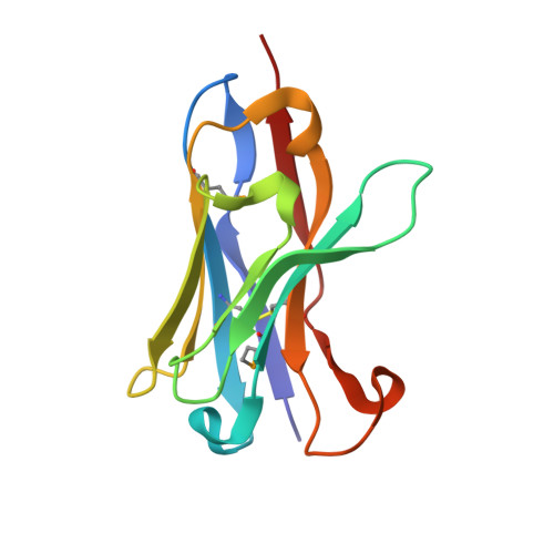
Crystal Structure of a Human Vh: Requirements for Maintaining a Monomeric Fragment

Dottorini, T.Vaughan, C.K.Walsh, M.A.Losurdo, P.Sollazzo, M.

(2004) Biochemistry 43: 622

  • DOI: https://doi.org/10.1021/bi035800b
  • Primary Citation of Related Structures:  
    1OL0

  • PubMed Abstract: 

    The variable domain of dromedary immunoglobulins comprises only the heavy chain and is missing the light-chain variable domain. This single domain is sufficient for antigen recognition and binding-half that required by other mammals. Human antibody-VHs have previously been camelized to be soluble stable fragments that retain antigen binding. Such engineered VHH are of interest in drug development, since they are nonimmunogenic, and in other biotechnology applications. We present the structure of a camelized human antibody fragment (cVH), which is a competitive and reversible inhibitor of the NS3 serine protease of the hepatitis C virus (HCV). In solution, this cVH undergoes a concentration-dependent monomer-dimer equilibrium. The structure confirms the minimum mutational requirements of the VL-binding face. The fragment also suggests a means by which the observed dimerization occurs, highlighting the importance of the composition of the CDR3 in maintaining a truly camelized VH.


  • Organizational Affiliation

    Istituto di Ricerche di Biologia Molecolare, Crystallography Unit, Via Pontina km 30600, 00040 Pomezia (Rm), Italy.


Macromolecules
Find similar proteins by:  (by identity cutoff)  |  3D Structure
Entity ID: 1
MoleculeChains Sequence LengthOrganismDetailsImage
IMMUNOGLOBULIN G
A, B
121Homo sapiensMutation(s): 0 
Entity Groups  
Sequence Clusters30% Identity50% Identity70% Identity90% Identity95% Identity100% Identity
Sequence Annotations
Expand
  • Reference Sequence
Small Molecules
Ligands 2 Unique
IDChains Name / Formula / InChI Key2D Diagram3D Interactions
SO4
Query on SO4

Download Ideal Coordinates CCD File 
C [auth A],
H [auth B],
I [auth B],
J [auth B],
K [auth B]
SULFATE ION
O4 S
QAOWNCQODCNURD-UHFFFAOYSA-L
GOL
Query on GOL

Download Ideal Coordinates CCD File 
D [auth A],
E [auth A],
F [auth A],
G [auth A],
L [auth B]
GLYCEROL
C3 H8 O3
PEDCQBHIVMGVHV-UHFFFAOYSA-N
Modified Residues  1 Unique
IDChains TypeFormula2D DiagramParent
MSE
Query on MSE
A, B
L-PEPTIDE LINKINGC5 H11 N O2 SeMET
Experimental Data & Validation

Experimental Data

  • Method: X-RAY DIFFRACTION
  • Resolution: 1.80 Å
  • R-Value Free: 0.180 
  • R-Value Work: 0.154 
  • R-Value Observed: 0.156 
  • Space Group: P 32 2 1
Unit Cell:
Length ( Å )Angle ( ˚ )
a = 75.3α = 90
b = 75.3β = 90
c = 101.78γ = 120
Software Package:
Software NamePurpose
REFMACrefinement
DENZOdata reduction
SCALEPACKdata scaling
AMoREphasing

Structure Validation

View Full Validation Report


Entry History 

Deposition Data

Revision History  (Full details and data files)

  • Version 1.0: 2004-01-22
    Type: Initial release
  • Version 1.1: 2011-05-08
    Changes: Version format compliance
  • Version 1.2: 2011-07-13
    Changes: Version format compliance
  • Version 1.3: 2019-05-08
    Changes: Data collection, Derived calculations, Experimental preparation, Other
  • Version 1.4: 2023-12-13
    Changes: Data collection, Database references, Other, Refinement description