1OJR

L-rhamnulose-1-phosphate aldolase from Escherichia coli (mutant E192A)


Experimental Data Snapshot

  • Method: X-RAY DIFFRACTION
  • Resolution: 1.35 Å
  • R-Value Free: 0.142 
  • R-Value Work: 0.111 
  • R-Value Observed: 0.112 

wwPDB Validation   3D Report Full Report


This is version 1.5 of the entry. See complete history


Literature

Structure and Catalytic Mechanism of L-Rhamnulose-1-Phosphate Aldolase.

Kroemer, M.Merkel, I.Schulz, G.E.

(2003) Biochemistry 42: 10560

  • DOI: https://doi.org/10.1021/bi0349266
  • Primary Citation of Related Structures:  
    1OJR

  • PubMed Abstract: 

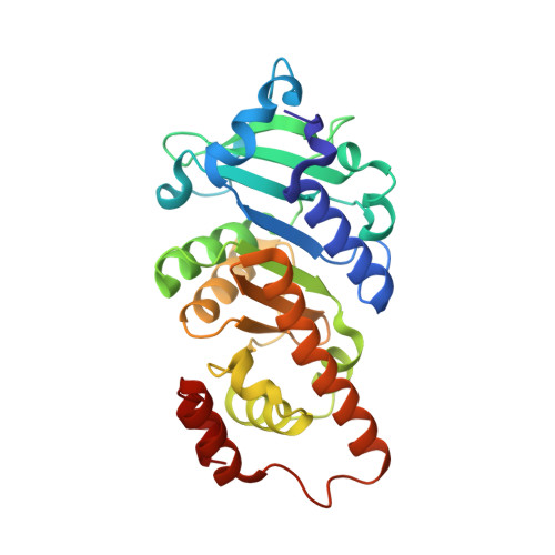
    The structure of L-rhamnulose-1-phosphate aldolase has been established at 1.35 A resolution in a crystal form that was obtained by a surface mutation and has one subunit of the C(4)-symmetric tetramer in the asymmetric unit. It confirms an earlier 2.7 A resolution structure which was determined in a complicated crystal form with 20 subunits per asymmetric unit. The chain fold and the active center are similar to those of L-fuculose-1-phosphate aldolase and L-ribulose-5-phosphate 4-epimerase. The active center similarity is supported by a structural comparison of all three enzymes and by the binding mode of the inhibitor phosphoglycolohydroxamate at the site of the product dihydroxyacetone phosphate for the two aldolases. The sensitivity of the catalytic rate to several mutations and a comparison with the established mechanism of the related aldolase give rise to a putative catalytic mechanism. This mechanism involves the same binding mode of the second product L-lactaldehyde in both aldolases, except for a 180 degrees flip of the aldehyde group distinguishing between the two epimers rhamnulose and fuculose. The N-terminal domain exhibits a correlated anisotropic mobility that channels the isotropic Brownian motion into a directed movement of the catalytic base and the substrate phosphate on the N-domain toward the zinc ion and the lactaldehyde on the C-terminal domain. We suggest that this movement supports the catalysis mechanically.


  • Organizational Affiliation

    Institut für Organische Chemie und Biochemie, Albertstrasse 21, 79104 Freiburg im Breisgau, Germany.


Macromolecules
Find similar proteins by:  (by identity cutoff)  |  3D Structure
Entity ID: 1
MoleculeChains Sequence LengthOrganismDetailsImage
RHAMNULOSE-1-PHOSPHATE ALDOLASE274Escherichia coliMutation(s): 1 
EC: 4.1.2.19
UniProt
Find proteins for P32169 (Escherichia coli (strain K12))
Explore P32169 
Go to UniProtKB:  P32169
Entity Groups  
Sequence Clusters30% Identity50% Identity70% Identity90% Identity95% Identity100% Identity
UniProt GroupP32169
Sequence Annotations
Expand
  • Reference Sequence
Small Molecules
Ligands 5 Unique
IDChains Name / Formula / InChI Key2D Diagram3D Interactions
PO4
Query on PO4

Download Ideal Coordinates CCD File 
C [auth A],
D [auth A]
PHOSPHATE ION
O4 P
NBIIXXVUZAFLBC-UHFFFAOYSA-K
GOL
Query on GOL

Download Ideal Coordinates CCD File 
G [auth A],
H [auth A],
I [auth A],
J [auth A]
GLYCEROL
C3 H8 O3
PEDCQBHIVMGVHV-UHFFFAOYSA-N
2HA
Query on 2HA

Download Ideal Coordinates CCD File 
E [auth A]Dihydroxyacetone
C3 H6 O3
RXKJFZQQPQGTFL-UHFFFAOYSA-N
DIO
Query on DIO

Download Ideal Coordinates CCD File 
F [auth A]1,4-DIETHYLENE DIOXIDE
C4 H8 O2
RYHBNJHYFVUHQT-UHFFFAOYSA-N
ZN
Query on ZN

Download Ideal Coordinates CCD File 
B [auth A]ZINC ION
Zn
PTFCDOFLOPIGGS-UHFFFAOYSA-N
Experimental Data & Validation

Experimental Data

  • Method: X-RAY DIFFRACTION
  • Resolution: 1.35 Å
  • R-Value Free: 0.142 
  • R-Value Work: 0.111 
  • R-Value Observed: 0.112 
  • Space Group: P 4 21 2
Unit Cell:
Length ( Å )Angle ( ˚ )
a = 108.024α = 90
b = 108.024β = 90
c = 57.166γ = 90
Software Package:
Software NamePurpose
REFMACrefinement
MOSFLMdata reduction
SCALAdata scaling
AMoREphasing

Structure Validation

View Full Validation Report


Entry History 

Deposition Data

Revision History  (Full details and data files)

  • Version 1.0: 2003-09-11
    Type: Initial release
  • Version 1.1: 2011-05-08
    Changes: Version format compliance
  • Version 1.2: 2011-07-13
    Changes: Version format compliance
  • Version 1.3: 2019-05-08
    Changes: Data collection, Experimental preparation, Other
  • Version 1.4: 2019-07-24
    Changes: Data collection
  • Version 1.5: 2023-12-13
    Changes: Data collection, Database references, Derived calculations, Other, Refinement description, Structure summary