1OHT

Peptidoglycan recognition protein LB


Experimental Data Snapshot

  • Method: X-RAY DIFFRACTION
  • Resolution: 2.00 Å
  • R-Value Free: 0.243 
  • R-Value Work: 0.216 
  • R-Value Observed: 0.216 

wwPDB Validation   3D Report Full Report


This is version 1.2 of the entry. See complete history


Literature

Crystal Structure of Peptidoglycan Recognition Protein Lb from Drosophila Melanogaster

Kim, M.-S.Byun, M.Oh, B.-H.

(2003) Nat Immunol 4: 787

  • DOI: https://doi.org/10.1038/ni952
  • Primary Citation of Related Structures:  
    1OHT

  • PubMed Abstract: 

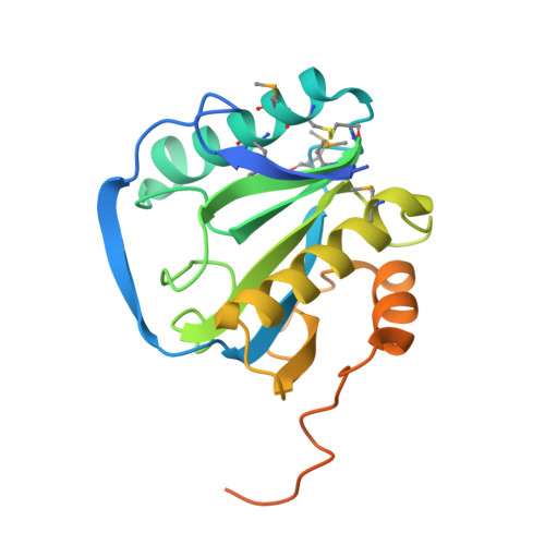
    The family of peptidoglycan recognition proteins (PGRPs) are associated with the recognition of the peptidoglycan of microbes and subsequent activation of signaling pathways for immune response. Here the crystal structure of Drosophila PGRP-LB is determined at a resolution of 2.0 A and shows an active-site cleft with a zinc cage. Poor conservation of surface residues at the cleft predicts a widely varying individual specificity of PGRPs for molecular patterns on microbial cell walls. At the back of this cleft is a putatively conserved distinctive groove. The location and mainly hydrophobic nature of the groove indicate that the back face serves for subsequent signaling after clustering of PGRP molecules by binding to polymeric cell wall components.


  • Organizational Affiliation

    Center for Biomolecular Recognition and Division of Molecular and Life Science, Department of Life Science, Pohang University of Science and Technology, Pohang, Kyungbuk, 790-784, Korea.


Macromolecules
Find similar proteins by:  (by identity cutoff)  |  3D Structure
Entity ID: 1
MoleculeChains Sequence LengthOrganismDetailsImage
CG14704 PROTEIN215Drosophila melanogasterMutation(s): 0 
UniProt
Find proteins for Q8INK6 (Drosophila melanogaster)
Explore Q8INK6 
Go to UniProtKB:  Q8INK6
Entity Groups  
Sequence Clusters30% Identity50% Identity70% Identity90% Identity95% Identity100% Identity
UniProt GroupQ8INK6
Sequence Annotations
Expand
  • Reference Sequence
Experimental Data & Validation

Experimental Data

  • Method: X-RAY DIFFRACTION
  • Resolution: 2.00 Å
  • R-Value Free: 0.243 
  • R-Value Work: 0.216 
  • R-Value Observed: 0.216 
  • Space Group: P 61 2 2
Unit Cell:
Length ( Å )Angle ( ˚ )
a = 40.561α = 90
b = 40.561β = 90
c = 337.997γ = 120
Software Package:
Software NamePurpose
CNSrefinement

Structure Validation

View Full Validation Report


Entry History 

Deposition Data

Revision History  (Full details and data files)

  • Version 1.0: 2003-07-18
    Type: Initial release
  • Version 1.1: 2011-05-08
    Changes: Version format compliance
  • Version 1.2: 2011-07-13
    Changes: Version format compliance