1O6T

Internalin (INLA, Listeria monocytogenes) - functional domain, uncomplexed


Experimental Data Snapshot

  • Method: X-RAY DIFFRACTION
  • Resolution: 1.60 Å
  • R-Value Free: 0.161 
  • R-Value Work: 0.131 
  • R-Value Observed: 0.133 

wwPDB Validation   3D Report Full Report


This is version 1.3 of the entry. See complete history


Literature

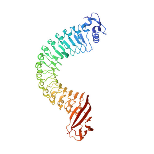
Structure of Internalin, a Major Invasion Protein of Listeria Monocytogenes, in Complex with its Human Receptor E-Cadherin

Schubert, W.-D.Urbanke, C.Ziehm, T.Beier, V.Machner, M.P.Domann, E.Wehland, J.Chakraborty, T.Heinz, D.W.

(2002) Cell 111: 825

  • DOI: https://doi.org/10.1016/s0092-8674(02)01136-4
  • Primary Citation of Related Structures:  
    1O6S, 1O6T, 1O6V

  • PubMed Abstract: 

    Listeria monocytogenes, a food-borne bacterial pathogen, enters mammalian cells by inducing its own phagocytosis. The listerial protein internalin (InlA) mediates bacterial adhesion and invasion of epithelial cells in the human intestine through specific interaction with its host cell receptor E-cadherin. We present the crystal structures of the functional domain of InlA alone and in a complex with the extracellular, N-terminal domain of human E-cadherin (hEC1). The leucine rich repeat (LRR) domain of InlA surrounds and specifically recognizes hEC1. Individual interactions were probed by mutagenesis and analytical ultracentrifugation. These include Pro16 of hEC1, a major determinant for human susceptibility to L. monocytogenes infection that is essential for intermolecular recognition. Our studies reveal the structural basis for host tro-pism of this bacterium and the molecular deception L. monocytogenes employs to exploit the E-cadherin system.


  • Organizational Affiliation

    Department of Structural Biology, German Research Center for Biotechnology (GBF), Mascheroder Weg 1, D-38124, Braunschweig, Germany.


Macromolecules
Find similar proteins by:  (by identity cutoff)  |  3D Structure
Entity ID: 1
MoleculeChains Sequence LengthOrganismDetailsImage
INTERNALIN A466Listeria monocytogenes EGD-eMutation(s): 0 
UniProt
Find proteins for P0DJM0 (Listeria monocytogenes serovar 1/2a (strain ATCC BAA-679 / EGD-e))
Explore P0DJM0 
Go to UniProtKB:  P0DJM0
Entity Groups  
Sequence Clusters30% Identity50% Identity70% Identity90% Identity95% Identity100% Identity
UniProt GroupP0DJM0
Sequence Annotations
Expand
  • Reference Sequence
Small Molecules
Ligands 5 Unique
IDChains Name / Formula / InChI Key2D Diagram3D Interactions
MES
Query on MES

Download Ideal Coordinates CCD File 
J [auth A],
K [auth A],
L [auth A],
M [auth A],
N [auth A]
2-(N-MORPHOLINO)-ETHANESULFONIC ACID
C6 H13 N O4 S
SXGZJKUKBWWHRA-UHFFFAOYSA-N
SO4
Query on SO4

Download Ideal Coordinates CCD File 
F [auth A],
G [auth A],
H [auth A],
I [auth A]
SULFATE ION
O4 S
QAOWNCQODCNURD-UHFFFAOYSA-L
CA
Query on CA

Download Ideal Coordinates CCD File 
C [auth A]CALCIUM ION
Ca
BHPQYMZQTOCNFJ-UHFFFAOYSA-N
CL
Query on CL

Download Ideal Coordinates CCD File 
D [auth A],
E [auth A]
CHLORIDE ION
Cl
VEXZGXHMUGYJMC-UHFFFAOYSA-M
MG
Query on MG

Download Ideal Coordinates CCD File 
B [auth A]MAGNESIUM ION
Mg
JLVVSXFLKOJNIY-UHFFFAOYSA-N
Experimental Data & Validation

Experimental Data

  • Method: X-RAY DIFFRACTION
  • Resolution: 1.60 Å
  • R-Value Free: 0.161 
  • R-Value Work: 0.131 
  • R-Value Observed: 0.133 
  • Space Group: C 1 2 1
Unit Cell:
Length ( Å )Angle ( ˚ )
a = 95.919α = 90
b = 55.881β = 113.07
c = 97.78γ = 90
Software Package:
Software NamePurpose
REFMACrefinement
DENZOdata reduction
SCALEPACKdata scaling
EPMRphasing

Structure Validation

View Full Validation Report


Entry History 

Deposition Data

Revision History  (Full details and data files)

  • Version 1.0: 2002-12-23
    Type: Initial release
  • Version 1.1: 2011-05-08
    Changes: Version format compliance
  • Version 1.2: 2011-07-13
    Changes: Version format compliance
  • Version 1.3: 2019-05-08
    Changes: Data collection, Derived calculations, Experimental preparation, Other