1NCO

STRUCTURE OF THE ANTITUMOR PROTEIN-CHROMOPHORE COMPLEX NEOCARZINOSTATIN


Experimental Data Snapshot

  • Method: X-RAY DIFFRACTION
  • Resolution: 1.80 Å
  • R-Value Observed: 0.199 

wwPDB Validation   3D Report Full Report


This is version 1.2 of the entry. See complete history


Literature

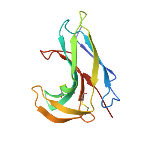
Crystal structure of neocarzinostatin, an antitumor protein-chromophore complex.

Kim, K.H.Kwon, B.M.Myers, A.G.Rees, D.C.

(1993) Science 262: 1042-1046

  • DOI: https://doi.org/10.1126/science.8235619
  • Primary Citation of Related Structures:  
    1NCO

  • PubMed Abstract: 

    Structures of the protein-chromophore complex and the apoprotein form of neocarzinostatin were determined at 1.8 angstrom resolution. Neocarzinostatin is composed of a labile chromophore with DNA-cleaving activity and a stabilizing protein. The chromophore displays marked nonlinearity of the triple bonds and is bound noncovalently in a pocket formed by the two protein domains. The chromophore pi-face interacts with the phenyl ring edges of Phe52 and Phe78. The amino sugar and carbonate groups of the chromophore are solvent exposed, whereas the epoxide, acetylene groups, and carbon C-12, the site of nucleophilic thiol addition during chromophore activation, are unexposed. The position of the amino group of the chromophore carbohydrate relative to C-12 supports the idea that the amino group plays a role in thiol activation.


  • Organizational Affiliation

    Division of Chemistry and Chemical Engineering, California Institute of Technology, Pasadena 91125.


Macromolecules
Find similar proteins by:  (by identity cutoff)  |  3D Structure
Entity ID: 1
MoleculeChains Sequence LengthOrganismDetailsImage
HOLO-NEOCARZINOSTATIN
A, B
113Streptomyces carzinostaticusMutation(s): 0 
UniProt
Find proteins for P0A3R9 (Streptomyces carzinostaticus)
Explore P0A3R9 
Go to UniProtKB:  P0A3R9
Entity Groups  
Sequence Clusters30% Identity50% Identity70% Identity90% Identity95% Identity100% Identity
UniProt GroupP0A3R9
Sequence Annotations
Expand
  • Reference Sequence
Small Molecules
Ligands 2 Unique
IDChains Name / Formula / InChI Key2D Diagram3D Interactions
CHR
Query on CHR

Download Ideal Coordinates CCD File 
D [auth B]NEOCARZINOSTATIN-CHROMOPHORE
C35 H33 N O12
QZGIWPZCWHMVQL-UIYAJPBUSA-N
MRD
Query on MRD

Download Ideal Coordinates CCD File 
C [auth A](4R)-2-METHYLPENTANE-2,4-DIOL
C6 H14 O2
SVTBMSDMJJWYQN-RXMQYKEDSA-N
Experimental Data & Validation

Experimental Data

  • Method: X-RAY DIFFRACTION
  • Resolution: 1.80 Å
  • R-Value Observed: 0.199 
  • Space Group: P 65
Unit Cell:
Length ( Å )Angle ( ˚ )
a = 90.4α = 90
b = 90.4β = 90
c = 52.5γ = 120
Software Package:
Software NamePurpose
X-PLORmodel building
TNTrefinement
X-PLORrefinement
X-PLORphasing

Structure Validation

View Full Validation Report


Entry History 

Deposition Data

Revision History  (Full details and data files)

  • Version 1.0: 1993-10-31
    Type: Initial release
  • Version 1.1: 2008-03-03
    Changes: Version format compliance
  • Version 1.2: 2011-07-13
    Changes: Version format compliance