1N8T

The crystal structure of phosphoglucose isomerase from rabbit muscle


Experimental Data Snapshot

  • Method: X-RAY DIFFRACTION
  • Resolution: 2.50 Å
  • R-Value Free: 0.234 
  • R-Value Work: 0.178 
  • R-Value Observed: 0.181 

wwPDB Validation   3D Report Full Report


This is version 1.4 of the entry. See complete history


Literature

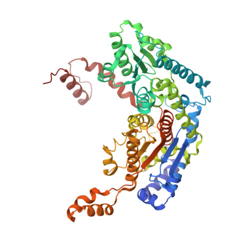
Structure of native phosphoglucose isomerase from rabbit: conformational changes associated with catalytic function.

Davies, C.Muirhead, H.

(2003) Acta Crystallogr D Biol Crystallogr 59: 453-465

  • DOI: https://doi.org/10.1107/s0907444902023387
  • Primary Citation of Related Structures:  
    1N8T

  • PubMed Abstract: 

    Phosphoglucose isomerase (PGI) is a housekeeping enzyme of metabolism that catalyses the interconversion of glucose 6-phosphate and fructose 6-phosphate, with roles in the glycolytic and gluconeogenic pathways. PGI is also a multifunctional protein that manifests the properties of a cytokine in a wide array of cellular processes, including the production of immunoglobulin by B cells and tumour-cell differentiation. The crystal structure of PGI in the native form from rabbit muscle has been solved at a resolution of 2.5 A by a combination of multiple isomorphous replacement and multi-crystal averaging techniques. Comparison with published structures of rabbit PGI in complex with three inhibitors and with the substrate fructose 6-phosphate reveals a number of conformational changes that may be associated with catalytic function. These occur in the small domain around the sugar phosphate-binding site, in a small helix carrying His388 and in a helix near the C-terminal end. One of these may be the structural rearrangement that has been postulated to be the rate-limiting step for catalysis.


  • Organizational Affiliation

    Department of Biochemistry and Molecular Biology, Medical University of South Carolina, Charleston, SC 29425, USA. davies@musc.edu


Macromolecules
Find similar proteins by:  (by identity cutoff)  |  3D Structure
Entity ID: 1
MoleculeChains Sequence LengthOrganismDetailsImage
Glucose-6-phosphate isomerase
A, B
557Oryctolagus cuniculusMutation(s): 0 
EC: 5.3.1.9
UniProt
Find proteins for Q9N1E2 (Oryctolagus cuniculus)
Explore Q9N1E2 
Go to UniProtKB:  Q9N1E2
Entity Groups  
Sequence Clusters30% Identity50% Identity70% Identity90% Identity95% Identity100% Identity
UniProt GroupQ9N1E2
Sequence Annotations
Expand
  • Reference Sequence
Experimental Data & Validation

Experimental Data

  • Method: X-RAY DIFFRACTION
  • Resolution: 2.50 Å
  • R-Value Free: 0.234 
  • R-Value Work: 0.178 
  • R-Value Observed: 0.181 
  • Space Group: C 2 2 21
Unit Cell:
Length ( Å )Angle ( ˚ )
a = 83.067α = 90
b = 115.174β = 90
c = 271.5γ = 90
Software Package:
Software NamePurpose
SCALEPACKdata scaling
MLPHAREphasing
REFMACrefinement

Structure Validation

View Full Validation Report


Entry History 

Deposition Data

Revision History  (Full details and data files)

  • Version 1.0: 2003-03-04
    Type: Initial release
  • Version 1.1: 2008-04-28
    Changes: Version format compliance
  • Version 1.2: 2011-07-13
    Changes: Version format compliance
  • Version 1.3: 2017-10-11
    Changes: Refinement description
  • Version 1.4: 2024-02-14
    Changes: Data collection, Database references