Determinants of Enzymatic Specificity in the Cys-Met-Metabolism PLP-Dependent Enzymes Family: Crystal Structure of Cystathionine gamma-lyase from Yeast and Intrafamiliar Structural Comparison
Messerschmidt, A., Worbs, M., Steegborn, C., Wahl, M.C., Huber, R., Laber, B., Clausen, T.(2003) Biol Chem 384: 373-386
- PubMed: 12715888
- DOI: https://doi.org/10.1515/BC.2003.043
- Primary Citation of Related Structures:
1N8P - PubMed Abstract:
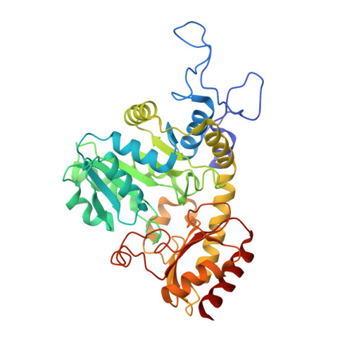
The crystal structure of cystathionine gamma-lyase (CGL) from yeast has been solved by molecular replacement at a resolution of 2.6 A. The molecule consists of 393 amino acid residues and one PLP moiety and is arranged in the crystal as a tetramer with D2 symmetry as in other related enzymes of the Cys-Met-metabolism PLP-dependent family like cystathionine beta-lyase (CBL). A structure comparison with other family members revealed surprising insights into the tuning of enzymatic specificity between the different family members. CGLs from yeast or human are virtually identical at their active sites to cystathionine gamma-synthase (CGS) from E. coli. Both CGLs and bacterial CGSs exhibit gamma-synthase and gamma-lyase activities depending on their position in the metabolic pathway and the available substrates. This group of enzymes has a glutamate (E333 in yeast CGL) which binds to the distal group of cystathionine (CTT) or the amino group of cysteine. Plant CGSs use homoserine phosphate instead of O-succinyl-homoserine as one substrate. This is reflected by a partially different active site structure in plant CGSs. In CGL and CBL the pseudosymmetric substrate must dock at the active site in different orientations, with S in gamma-position (CBL) or in delta-position (CGL). The conserved glutamate steers the substrate as seen in other CGLs. In CBLs this position is occupied by either tyrosine or hydrophobic residues directing binding of CTT such that S is in the in gamma-position. In methionine gamma-lyase a hydrophic patch operates as recognition site for the methyl group of the methionine substrate.
Organizational Affiliation:
Max-Planck-Institut for Biochemie, Abteilung Strukturforschung, Am Klopferspitz 18a, D-82152 Planegg-Martinsried, Germany.