1MZ4

Crystal Structure of Cytochrome c550 from Thermosynechococcus elongatus


Experimental Data Snapshot

  • Method: X-RAY DIFFRACTION
  • Resolution: 1.80 Å
  • R-Value Free: 0.202 
  • R-Value Work: 0.182 
  • R-Value Observed: 0.182 

wwPDB Validation   3D Report Full Report


Ligand Structure Quality Assessment 


This is version 2.0 of the entry. See complete history


Literature

Structural and EPR characterization of the soluble form of cytochrome c-550 and of the psbV2 gene product from the cyanobacterium Thermosynechococcus elongatus.

Kerfeld, C.A.Sawaya, M.R.Bottin, H.Tran, K.T.Sugiura, M.Cascio, D.Desbois, A.Yeates, T.O.Kirilovsky, D.Boussac, A.

(2003) Plant Cell Physiol 44: 697-706

  • DOI: https://doi.org/10.1093/pcp/pcg084
  • Primary Citation of Related Structures:  
    1MZ4

  • PubMed Abstract: 

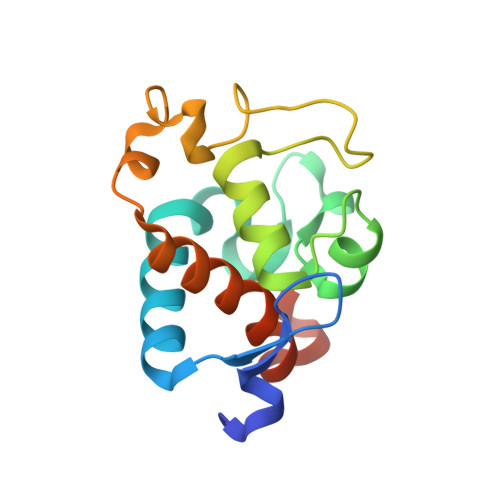
    First, the crystal structure of cytochrome c-550 (the psbV1 gene product) from the thermophilic cyanobacterium Thermosynechococcus elongatus has been determined to a resolution of 1.8 A. A comparison of the T. elongatus cytochrome c-550 structure to its counterparts from mesophilic organisms, Synechocystis 6803 and Arthrospira maxima, suggests that increased numbers of hydrogen bonds may play a role in the structural basis of thermostability. The cytochrome c-550 in T. elongatus also differs from that in Synechocystis 6803 and Arthrospira maxima in its lack of dimerization and the presence of a trigonal planar molecule, possibly bicarbonate, tightly bound to the heme propionate oxygen atoms. Cytochromes c-550 from T. elongatus, Synechocystis 6803 and Arthrospira maxima exhibit different EPR spectra. A correlation has been done between the heme-axial ligands geometries and the rhombicity calculated from the EPR spectra. This correlation indicates that binding of cytochrome c-550 to Photosystem II is accompanied by structural changes in the heme vicinity. Second, the psbV2 gene product has been found and purified. The UV-visible, EPR and Raman spectra are reported. From the spectroscopic data and from a theoretical structural model based on the cytochrome c-550 structure it is proposed that the 6th ligand of the heme-iron is the Tyr86.


  • Organizational Affiliation

    Molecular Biology Institute, UCLA, Box 951570, Los Angeles, CA 90095-1570, USA. kerfeld@mbi.ucla.edu


Macromolecules
Find similar proteins by:  (by identity cutoff)  |  3D Structure
Entity ID: 1
MoleculeChains Sequence LengthOrganismDetailsImage
cytochrome c550137Thermosynechococcus vestitusMutation(s): 0 
UniProt
Find proteins for P0A386 (Thermosynechococcus vestitus (strain NIES-2133 / IAM M-273 / BP-1))
Explore P0A386 
Go to UniProtKB:  P0A386
Entity Groups  
Sequence Clusters30% Identity50% Identity70% Identity90% Identity95% Identity100% Identity
UniProt GroupP0A386
Sequence Annotations
Expand
  • Reference Sequence
Small Molecules
Ligands 4 Unique
IDChains Name / Formula / InChI Key2D Diagram3D Interactions
HEC
Query on HEC

Download Ideal Coordinates CCD File 
H [auth A]HEME C
C34 H34 Fe N4 O4
HXQIYSLZKNYNMH-LJNAALQVSA-N
PO4
Query on PO4

Download Ideal Coordinates CCD File 
B [auth A],
C [auth A],
D [auth A],
E [auth A],
F [auth A]
PHOSPHATE ION
O4 P
NBIIXXVUZAFLBC-UHFFFAOYSA-K
GOL
Query on GOL

Download Ideal Coordinates CCD File 
I [auth A],
J [auth A],
K [auth A],
L [auth A],
M [auth A]
GLYCEROL
C3 H8 O3
PEDCQBHIVMGVHV-UHFFFAOYSA-N
BCT
Query on BCT

Download Ideal Coordinates CCD File 
G [auth A]BICARBONATE ION
C H O3
BVKZGUZCCUSVTD-UHFFFAOYSA-M
Experimental Data & Validation

Experimental Data

  • Method: X-RAY DIFFRACTION
  • Resolution: 1.80 Å
  • R-Value Free: 0.202 
  • R-Value Work: 0.182 
  • R-Value Observed: 0.182 
  • Space Group: P 32 2 1
Unit Cell:
Length ( Å )Angle ( ˚ )
a = 69.641α = 90
b = 69.641β = 90
c = 92.849γ = 120
Software Package:
Software NamePurpose
DENZOdata reduction
SCALEPACKdata scaling
MLPHAREphasing
CNSrefinement

Structure Validation

View Full Validation Report


Ligand Structure Quality Assessment 


Entry History 

Deposition Data

Revision History  (Full details and data files)

  • Version 1.0: 2003-09-23
    Type: Initial release
  • Version 1.1: 2007-10-16
    Changes: Version format compliance
  • Version 1.2: 2011-07-13
    Changes: Non-polymer description, Version format compliance
  • Version 2.0: 2021-03-03
    Changes: Atomic model, Data collection, Derived calculations, Non-polymer description, Structure summary