1MUQ

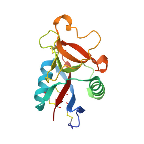
X-ray Crystal Structure of Rattlesnake Venom Complexed With Thiodigalactoside


Experimental Data Snapshot

  • Method: X-RAY DIFFRACTION
  • Resolution: 2.30 Å
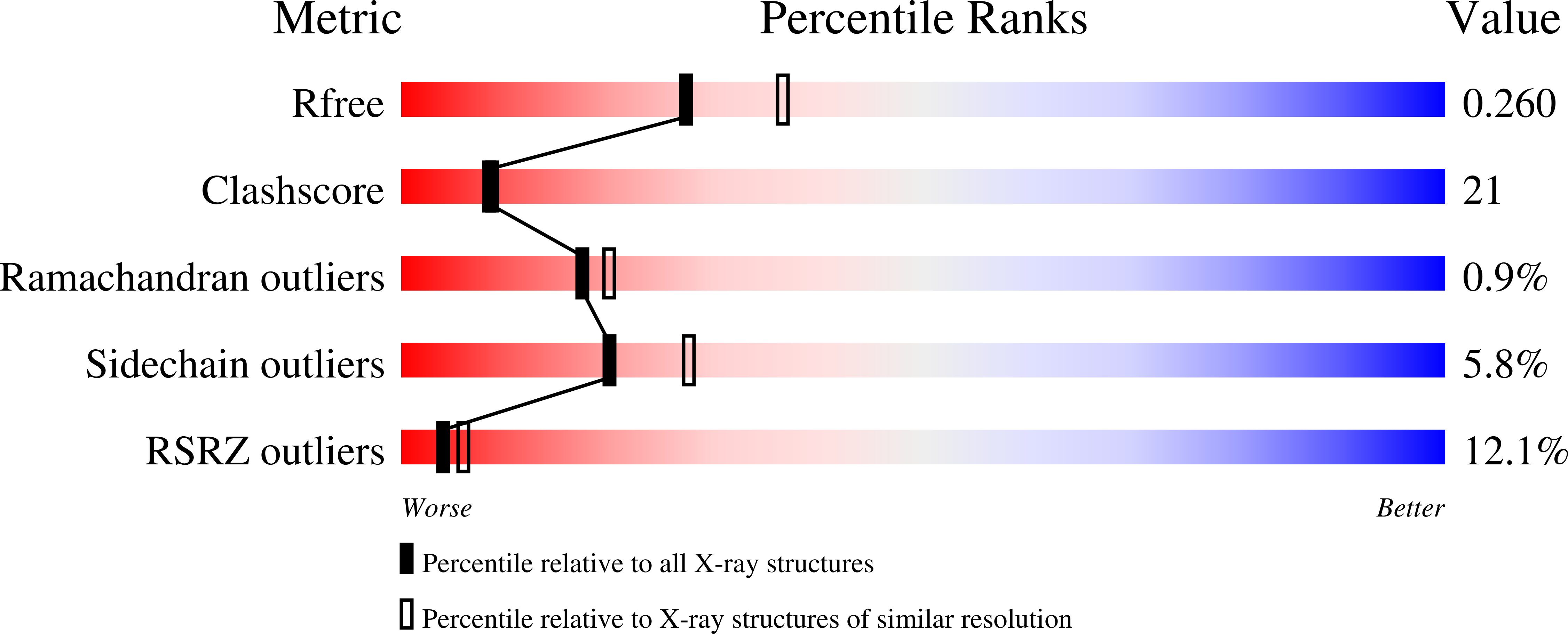
  • R-Value Free: 0.257 
  • R-Value Work: 0.210 
  • R-Value Observed: 0.210 

wwPDB Validation   3D Report Full Report


Ligand Structure Quality Assessment 


This is version 3.0 of the entry. See complete history


Literature

X-ray Crystal Structure of a Galactose-Specific C-Type Lectin Possessing a Novel Decameric Quaternary Structure.

Walker, J.R.Nagar, B.Young, N.M.Hirama, T.Rini, J.M.

(2004) Biochemistry 43: 3783-3792

  • DOI: https://doi.org/10.1021/bi035871a
  • Primary Citation of Related Structures:  
    1JZN, 1MUQ

  • PubMed Abstract: 

    Rattlesnake venom lectin (RSL) from the western diamondback rattlesnake (Crotalus atrox) is an oligomeric galactose-specific C-type lectin. The X-ray crystal structure of RSL, in complex with lactose and thiodigalactoside, at 2.2 and 2.3 A resolution, respectively, reveals a decameric protein composed of two 5-fold symmetric pentamers arranged in a staggered, back-to-back orientation. Each monomer corresponds to a single canonical C-type lectin carbohydrate recognition domain devoid of accessory domains and is disulfide-bonded to a monomer in the other pentamer. The structure is the first example of that of a carbohydrate complex of a vertebrate galactose-specific C-type lectin. The 10 carbohydrate-binding sites, located on the rim of the decamer, suggest a role for multivalent interactions and a mechanism for RSL's ability to promote receptor cross-linking and cell aggregation.


  • Organizational Affiliation

    Department of Molecular and Medical Genetics, University of Toronto, Toronto, Ontario M5S 1A8, Canada.


Macromolecules
Find similar proteins by:  (by identity cutoff)  |  3D Structure
Entity ID: 1
MoleculeChains Sequence LengthOrganismDetailsImage
Galactose-specific lectin
A, B, C, D, E
135Crotalus atroxMutation(s): 0 
UniProt
Find proteins for P21963 (Crotalus atrox)
Explore P21963 
Go to UniProtKB:  P21963
Entity Groups  
Sequence Clusters30% Identity50% Identity70% Identity90% Identity95% Identity100% Identity
UniProt GroupP21963
Sequence Annotations
Expand
  • Reference Sequence
Oligosaccharides

Help

Entity ID: 2
MoleculeChains Length2D Diagram Glycosylation3D Interactions
1-thio-beta-D-galactopyranose-(1-1)-beta-D-galactopyranoseF [auth G]2N/AN/A
Glycosylation Resources
GlyTouCan:  G61532BD
GlyCosmos:  G61532BD
Small Molecules
Ligands 3 Unique
IDChains Name / Formula / InChI Key2D Diagram3D Interactions
GAL
Query on GAL

Download Ideal Coordinates CCD File 
I [auth A],
N [auth C],
S [auth E]
beta-D-galactopyranose
C6 H12 O6
WQZGKKKJIJFFOK-FPRJBGLDSA-N
CA
Query on CA

Download Ideal Coordinates CCD File 
G [auth A],
J [auth B],
L [auth C],
O [auth D],
Q [auth E]
CALCIUM ION
Ca
BHPQYMZQTOCNFJ-UHFFFAOYSA-N
NA
Query on NA

Download Ideal Coordinates CCD File 
H [auth A],
K [auth B],
M [auth C],
P [auth D],
R [auth E]
SODIUM ION
Na
FKNQFGJONOIPTF-UHFFFAOYSA-N
Biologically Interesting Molecules (External Reference) 1 Unique
Experimental Data & Validation

Experimental Data

  • Method: X-RAY DIFFRACTION
  • Resolution: 2.30 Å
  • R-Value Free: 0.257 
  • R-Value Work: 0.210 
  • R-Value Observed: 0.210 
  • Space Group: C 2 2 21
Unit Cell:
Length ( Å )Angle ( ˚ )
a = 110.454α = 90
b = 151.234β = 90
c = 95.014γ = 90
Software Package:
Software NamePurpose
DENZOdata reduction
SCALEPACKdata scaling
PHASESphasing
CNSrefinement

Structure Validation

View Full Validation Report


Ligand Structure Quality Assessment 


Entry History 

Deposition Data

Revision History  (Full details and data files)

  • Version 1.0: 2003-07-01
    Type: Initial release
  • Version 1.1: 2008-04-28
    Changes: Version format compliance
  • Version 1.2: 2011-07-13
    Changes: Derived calculations, Version format compliance
  • Version 2.0: 2020-07-29
    Type: Remediation
    Reason: Carbohydrate remediation
    Changes: Advisory, Atomic model, Data collection, Derived calculations, Non-polymer description, Structure summary
  • Version 3.0: 2020-10-14
    Changes: Atomic model, Data collection, Derived calculations, Structure summary