1MD7

Monomeric structure of the zymogen of complement protease C1r


Experimental Data Snapshot

  • Method: X-RAY DIFFRACTION
  • Resolution: 3.20 Å
  • R-Value Free: 0.285 
  • R-Value Work: 0.217 
  • R-Value Observed: 0.243 

wwPDB Validation   3D Report Full Report


Ligand Structure Quality Assessment 


This is version 2.1 of the entry. See complete history


Literature

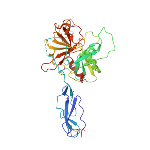
Monomeric structures of the zymogen and active catalytic domain of complement protease c1r: further insights into the c1 activation mechanism

Budayova-Spano, M.Grabarse, W.Thielens, N.M.Hillen, H.Lacroix, M.Schmidt, M.Fontecilla-Camps, J.Arlaud, G.J.Gaboriaud, C.

(2002) Structure 10: 1509-1519

  • DOI: https://doi.org/10.1016/s0969-2126(02)00881-x
  • Primary Citation of Related Structures:  
    1MD7, 1MD8

  • PubMed Abstract: 

    C1r is the serine protease (SP) that mediates autoactivation of C1, the complex that triggers the classical complement pathway. We have determined the crystal structure of two fragments from the human C1r catalytic domain, each encompassing the second complement control protein (CCP2) module and the SP domain. The wild-type species has an active structure, whereas the S637A mutant is a zymogen. The structures reveal a restricted hinge flexibility of the CCP2-SP interface, and both are characterized by the unique alpha-helical conformation of loop E. The zymogen activation domain exhibits high mobility, and the active structure shows a restricted access to most substrate binding subsites. Further implications relevant to the C1r self-activation process are derived from protein-protein interactions in the crystals.


  • Organizational Affiliation

    LCCP, Institut de Biologie Structurale Jean-Pierre Ebel, CEA-CNRS, 41 rue Jules Horowitz, Grenoble, France.


Macromolecules
Find similar proteins by:  (by identity cutoff)  |  3D Structure
Entity ID: 1
MoleculeChains Sequence LengthOrganismDetailsImage
C1R COMPLEMENT SERINE PROTEASE328Homo sapiensMutation(s): 1 
EC: 3.4.21.41
UniProt & NIH Common Fund Data Resources
Find proteins for P00736 (Homo sapiens)
Explore P00736 
Go to UniProtKB:  P00736
PHAROS:  P00736
Entity Groups  
Sequence Clusters30% Identity50% Identity70% Identity90% Identity95% Identity100% Identity
UniProt GroupP00736
Sequence Annotations
Expand
  • Reference Sequence
Small Molecules
Ligands 1 Unique
IDChains Name / Formula / InChI Key2D Diagram3D Interactions
NAG
Query on NAG

Download Ideal Coordinates CCD File 
B [auth A]2-acetamido-2-deoxy-beta-D-glucopyranose
C8 H15 N O6
OVRNDRQMDRJTHS-FMDGEEDCSA-N
Experimental Data & Validation

Experimental Data

  • Method: X-RAY DIFFRACTION
  • Resolution: 3.20 Å
  • R-Value Free: 0.285 
  • R-Value Work: 0.217 
  • R-Value Observed: 0.243 
  • Space Group: P 64
Unit Cell:
Length ( Å )Angle ( ˚ )
a = 167.06α = 90
b = 167.06β = 90
c = 43.648γ = 120
Software Package:
Software NamePurpose
MOSFLMdata reduction
SCALAdata scaling
AMoREphasing
CNSrefinement
CCP4data scaling

Structure Validation

View Full Validation Report


Ligand Structure Quality Assessment 


Entry History 

Deposition Data

Revision History  (Full details and data files)

  • Version 1.0: 2003-08-07
    Type: Initial release
  • Version 1.1: 2008-04-28
    Changes: Version format compliance
  • Version 1.2: 2011-07-13
    Changes: Version format compliance
  • Version 2.0: 2020-07-29
    Type: Remediation
    Reason: Carbohydrate remediation
    Changes: Advisory, Atomic model, Data collection, Derived calculations, Non-polymer description, Structure summary
  • Version 2.1: 2021-11-10
    Changes: Advisory, Database references, Structure summary