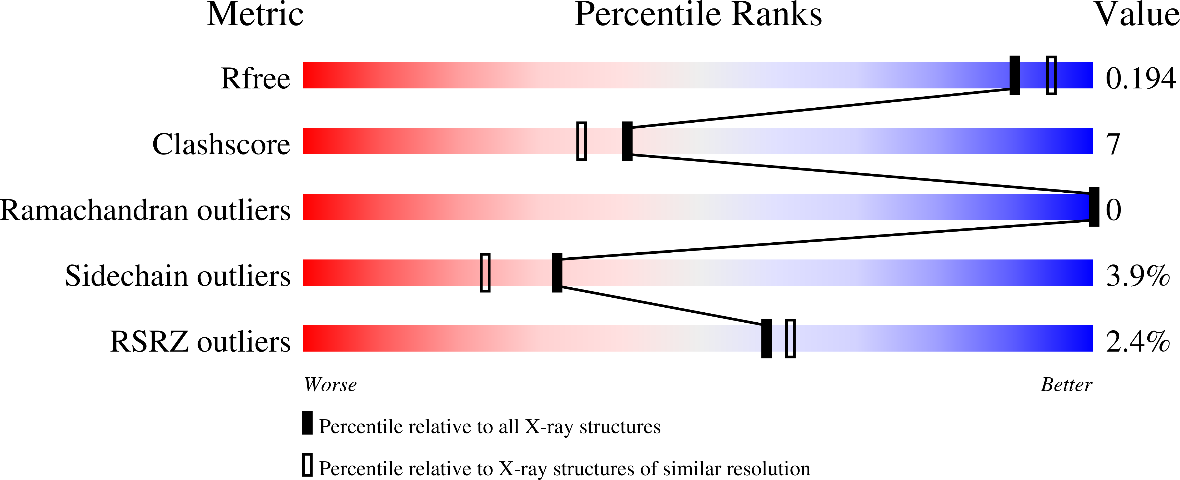
1M8S

Crystal Structures of Cadmium-binding Acidic Phospholipase A2 from the Venom of Agkistrodon halys pallas at 1.9 Resolution (crystal grown at pH 5.9)


Experimental Data Snapshot

  • Method: X-RAY DIFFRACTION
  • Resolution: 1.90 Å
  • R-Value Free: 0.209 
  • R-Value Work: 0.202 
  • R-Value Observed: 0.202 

wwPDB Validation   3D Report Full Report


This is version 1.3 of the entry. See complete history


Literature

Structures of cadmium-binding acidic phospholipase A(2) from the venom of Agkistrodon halys Pallas at 1.9A resolutio

Xu, S.Gu, L.Jiang, T.Zhou, Y.Lin, Z.

(2003) Biochem Biophys Res Commun 300: 271-277

  • DOI: https://doi.org/10.1016/s0006-291x(02)02833-4
  • Primary Citation of Related Structures:  
    1M8R, 1M8S

  • PubMed Abstract: 

    Phospholipase A(2) coordinates Ca(2+) ion through three carbonyl oxygen atoms of residues 28, 30, and 32, two carboxyl oxygen atoms of residue Asp49, and two (or one) water molecules, forming seven (or six) coordinate geometry of Ca(2+) ligands. Two crystal structures of cadmium-binding acidic phospholipase A(2) from the venom of Agkistrodon halys Pallas (i.e., Agkistrodon blomhoffii brevicaudus) at different pH values (5.9 and 7.4) were determined to 1.9A resolution by the isomorphous difference Fourier method. The well-refined structures revealed that a Cd(2+) ion occupied the position expected for a Ca(2+) ion, and that the substitution of Cd(2+) for Ca(2+) resulted in detectable changes in the metal-binding region: one of the carboxyl oxygen atoms from residue Asp49 was farther from the metal ion while the other one was closer and there were no water molecules coordinating to the metal ion. Thus the Cd(2+)-binding region appears to have four coordinating oxygen ligands. The cadmium binding to the enzyme induced no other significant conformational change in the enzyme molecule elsewhere. The mechanism for divalent cadmium cation to support substrate binding but not catalysis is discussed.


  • Organizational Affiliation

    National Laboratory of Biomacromolecules, Institute of Biophysics, Chinese Academy of Sciences, Beijing 100101, China.


Macromolecules
Find similar proteins by:  (by identity cutoff)  |  3D Structure
Entity ID: 1
MoleculeChains Sequence LengthOrganismDetailsImage
phospholipase a2124Gloydius halysMutation(s): 0 
EC: 3.1.1.4
UniProt
Find proteins for O42191 (Gloydius halys)
Explore O42191 
Go to UniProtKB:  O42191
Entity Groups  
Sequence Clusters30% Identity50% Identity70% Identity90% Identity95% Identity100% Identity
UniProt GroupO42191
Sequence Annotations
Expand
  • Reference Sequence
Experimental Data & Validation

Experimental Data

  • Method: X-RAY DIFFRACTION
  • Resolution: 1.90 Å
  • R-Value Free: 0.209 
  • R-Value Work: 0.202 
  • R-Value Observed: 0.202 
  • Space Group: P 61
Unit Cell:
Length ( Å )Angle ( ˚ )
a = 83.16α = 90
b = 83.16β = 90
c = 32.78γ = 120
Software Package:
Software NamePurpose
DENZOdata reduction
SCALEPACKdata scaling
CNSrefinement
CNSphasing

Structure Validation

View Full Validation Report


Entry History 

Deposition Data

Revision History  (Full details and data files)

  • Version 1.0: 2003-02-11
    Type: Initial release
  • Version 1.1: 2008-04-28
    Changes: Version format compliance
  • Version 1.2: 2011-07-13
    Changes: Version format compliance
  • Version 1.3: 2023-10-25
    Changes: Data collection, Database references, Derived calculations, Refinement description