


Structural Snapshots in the Molecular Mechanism of PFO Revealed
Rossjohn, J., Parker, M., Polekhina, G., Feil, S., Tweten, R.To be published.
Experimental Data Snapshot
wwPDB Validation 3D Report Full Report

Entity ID: 1 | |||||
|---|---|---|---|---|---|
| Molecule | Chains | Sequence Length | Organism | Details | Image |
| perfringolysin O | 471 | Clostridium perfringens | Mutation(s): 0 Gene Names: pfoa Membrane Entity: Yes | 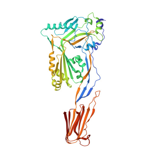 | |
UniProt | |||||
Find proteins for P0C2E9 (Clostridium perfringens (strain 13 / Type A)) Explore P0C2E9 Go to UniProtKB: P0C2E9 | |||||
Entity Groups | |||||
| Sequence Clusters | 30% Identity50% Identity70% Identity90% Identity95% Identity100% Identity | ||||
| UniProt Group | P0C2E9 | ||||
Sequence AnnotationsExpand | |||||
| |||||
| Length ( Å ) | Angle ( ˚ ) |
|---|---|
| a = 130.41 | α = 90 |
| b = 130.41 | β = 90 |
| c = 129.9 | γ = 120 |
| Software Name | Purpose |
|---|---|
| DENZO | data reduction |
| SCALEPACK | data scaling |
| AMoRE | phasing |
| CNS | refinement |
RCSB PDB (citation) is hosted by
RCSB PDB is a member of the

