1LOD

INTERACTION OF A LEGUME LECTIN WITH TWO COMPONENTS OF THE BACTERIAL CELL WALL


Experimental Data Snapshot

  • Method: X-RAY DIFFRACTION
  • Resolution: 2.05 Å
  • R-Value Work: 0.197 
  • R-Value Observed: 0.197 

wwPDB Validation   3D Report Full Report


Ligand Structure Quality Assessment 


This is version 1.4 of the entry. See complete history


Literature

Interaction of a legume lectin with two components of the bacterial cell wall. A crystallographic study.

Bourne, Y.Ayouba, A.Rouge, P.Cambillau, C.

(1994) J Biol Chem 269: 9429-9435

  • Primary Citation of Related Structures:  
    1LOC, 1LOD

  • PubMed Abstract: 

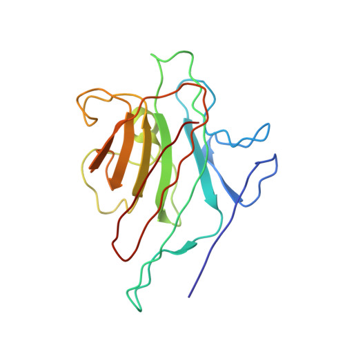
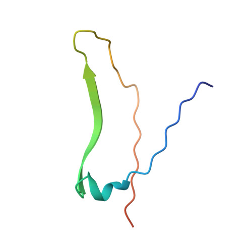
    We describe herein the refined high resolution x-ray structures of two components of the bacterial cell wall, muramic acid and muramyl dipeptide complexed to isolectin I from Lathyrus ochrus seeds. In both complexes, only the ring hydroxyl oxygen atoms of the bound sugar establish direct hydrogen bonds with isolectin I, as in the case of all the previously determined monosaccharide-lectin complexes. In addition, the lactyl methyl of both components strongly interacts via hydrophobic contacts with the side chains of residues Tyr100 and Trp128 of isolectin I, which could explain the higher affinity of isolectin I for muramic acid as compared with glucose. These 2 residues, however, are not involved in the stabilization of the oligosaccharide-isolectin I complexes. The dipeptide (D-Ala-D-iGln) of the second component is in stacking interaction with the N-acetyl group of glucose and with loop Gly97-Gly98 of isolectin I. In addition to these van der Waals' contacts, the dipeptide interacts with the lectin via well ordered water molecules also. Superposition of the structures of the muramyl dipeptide complex and of the muramic acid complex shows that the glucose ring in the dipeptide compound is tilted by about 15 degrees in comparison with that of muramic acid. The fact that the lactyl group has the same confrontation in both components reveals that the lectin is stereospecific and recognizes only diastereoisomer S of this group, which better fits the saccharide-binding site.


  • Organizational Affiliation

    Laboratorie de Cristallographie et de Cristallisation des Macromolécules Biologiques, Centre National de la Recherche Scientifique Unité de Recherche associée 1296, Faculté de Médecine Secteur-Nord, Marseille, France.


Macromolecules
Find similar proteins by:  (by identity cutoff)  |  3D Structure
Entity ID: 1
MoleculeChains Sequence LengthOrganismDetailsImage
LEGUME ISOLECTIN I (ALPHA CHAIN)
A, C, E, G
181Lathyrus ochrusMutation(s): 0 
UniProt
Find proteins for P04122 (Lathyrus ochrus)
Explore P04122 
Go to UniProtKB:  P04122
Entity Groups  
Sequence Clusters30% Identity50% Identity70% Identity90% Identity95% Identity100% Identity
UniProt GroupP04122
Sequence Annotations
Expand
  • Reference Sequence
Find similar proteins by:  (by identity cutoff)  |  3D Structure
Entity ID: 2
MoleculeChains Sequence LengthOrganismDetailsImage
LEGUME ISOLECTIN I (BETA CHAIN)
B, D, F, H
52Lathyrus ochrusMutation(s): 0 
UniProt
Find proteins for P12306 (Lathyrus ochrus)
Explore P12306 
Go to UniProtKB:  P12306
Entity Groups  
Sequence Clusters30% Identity50% Identity70% Identity90% Identity95% Identity100% Identity
UniProt GroupP12306
Sequence Annotations
Expand
  • Reference Sequence
Small Molecules
Ligands 3 Unique
IDChains Name / Formula / InChI Key2D Diagram3D Interactions
MUR
Query on MUR

Download Ideal Coordinates CCD File 
K [auth A],
N [auth C],
Q [auth E],
T [auth G]
beta-muramic acid
C9 H17 N O7
MSFSPUZXLOGKHJ-KTZFPWNASA-N
MN
Query on MN

Download Ideal Coordinates CCD File 
J [auth A],
M [auth C],
P [auth E],
S [auth G]
MANGANESE (II) ION
Mn
WAEMQWOKJMHJLA-UHFFFAOYSA-N
CA
Query on CA

Download Ideal Coordinates CCD File 
I [auth A],
L [auth C],
O [auth E],
R [auth G]
CALCIUM ION
Ca
BHPQYMZQTOCNFJ-UHFFFAOYSA-N
Experimental Data & Validation

Experimental Data

  • Method: X-RAY DIFFRACTION
  • Resolution: 2.05 Å
  • R-Value Work: 0.197 
  • R-Value Observed: 0.197 
  • Space Group: P 1 21 1
Unit Cell:
Length ( Å )Angle ( ˚ )
a = 56.4α = 90
b = 139.8β = 91
c = 62.7γ = 90
Software Package:
Software NamePurpose
X-PLORmodel building
X-PLORrefinement
X-PLORphasing

Structure Validation

View Full Validation Report


Ligand Structure Quality Assessment 


Entry History 

Deposition Data

Revision History  (Full details and data files)

  • Version 1.0: 1994-04-30
    Type: Initial release
  • Version 1.1: 2008-03-24
    Changes: Version format compliance
  • Version 1.2: 2011-07-13
    Changes: Derived calculations, Version format compliance
  • Version 1.3: 2020-07-29
    Type: Remediation
    Reason: Carbohydrate remediation
    Changes: Advisory, Data collection, Database references, Derived calculations, Other, Structure summary
  • Version 1.4: 2024-02-14
    Changes: Data collection, Database references, Structure summary