1LHR

Crystal Structure of Pyridoxal Kinase complexed with ATP


Experimental Data Snapshot

  • Method: X-RAY DIFFRACTION
  • Resolution: 2.60 Å
  • R-Value Free: 0.222 
  • R-Value Work: 0.193 
  • R-Value Observed: 0.198 

wwPDB Validation   3D Report Full Report


Ligand Structure Quality Assessment 


This is version 1.3 of the entry. See complete history


Literature

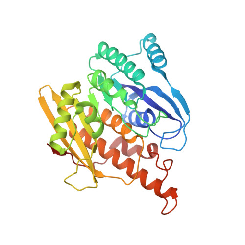
Crystal structure of brain pyridoxal kinase, a novel member of the ribokinase superfamily

Li, M.H.Kwok, F.Chang, W.R.Lau, C.K.Zhang, J.P.Lo, S.C.Jiang, T.Liang, D.C.

(2002) J Biol Chem 277: 46385-46390

  • DOI: https://doi.org/10.1074/jbc.M208600200
  • Primary Citation of Related Structures:  
    1LHP, 1LHR

  • PubMed Abstract: 

    The three-dimensional structures of brain pyridoxal kinase and its complex with the nucleotide ATP have been elucidated in the dimeric form at 2.1 and 2.6 A, respectively. Results have shown that pyridoxal kinase, as an enzyme obeying random sequential kinetics in catalysis, does not possess a lid shape structure common to all kinases in the ribokinase superfamily. This finding has been shown to be in line with the condition that pyridoxal kinase binds substrates with variable sizes of chemical groups at position 4 of vitamin B(6) and its derivatives. In addition, the enzyme contains a 12-residue peptide loop in the active site for the prevention of premature hydrolysis of ATP. Conserved amino acid residues Asp(118) and Tyr(127) in the peptide loop could be moved to a position covering the nucleotide after its binding so that its chance to hydrolyze in the aqueous environment of the active site was reduced. With respect to the evolutionary trend of kinase enzymes, the existence of this loop in pyridoxal kinase could be classified as an independent category in the ribokinase superfamily according to the structural feature found and mechanism followed in catalysis.


  • Organizational Affiliation

    National Laboratory of Biomacromolecules, Institute of Biophysics, Chinese Academy of Science, Beijing 100101, China.


Macromolecules
Find similar proteins by:  (by identity cutoff)  |  3D Structure
Entity ID: 1
MoleculeChains Sequence LengthOrganismDetailsImage
Pyridoxal kinase
A, B
312Ovis ariesMutation(s): 0 
EC: 2.7.1.35
UniProt
Find proteins for P82197 (Ovis aries)
Explore P82197 
Go to UniProtKB:  P82197
Entity Groups  
Sequence Clusters30% Identity50% Identity70% Identity90% Identity95% Identity100% Identity
UniProt GroupP82197
Sequence Annotations
Expand
  • Reference Sequence
Experimental Data & Validation

Experimental Data

  • Method: X-RAY DIFFRACTION
  • Resolution: 2.60 Å
  • R-Value Free: 0.222 
  • R-Value Work: 0.193 
  • R-Value Observed: 0.198 
  • Space Group: P 21 21 21
Unit Cell:
Length ( Å )Angle ( ˚ )
a = 59α = 90
b = 93.8β = 90
c = 128.8γ = 90
Software Package:
Software NamePurpose
DENZOdata reduction
SCALEPACKdata scaling
CNSrefinement
CNSphasing

Structure Validation

View Full Validation Report


Ligand Structure Quality Assessment 


Entry History 

Deposition Data

Revision History  (Full details and data files)

  • Version 1.0: 2003-02-11
    Type: Initial release
  • Version 1.1: 2008-04-28
    Changes: Version format compliance
  • Version 1.2: 2011-07-13
    Changes: Version format compliance
  • Version 1.3: 2024-03-13
    Changes: Data collection, Database references, Derived calculations