1L2L

Crystal structure of ADP-dependent glucokinase from a Pyrococcus Horikoshii


Experimental Data Snapshot

  • Method: X-RAY DIFFRACTION
  • Resolution: 2.00 Å
  • R-Value Free: 0.277 
  • R-Value Work: 0.213 
  • R-Value Observed: 0.213 

wwPDB Validation   3D Report Full Report


This is version 1.3 of the entry. See complete history


Literature

Crystal structure of the ADP-dependent glucokinase from Pyrococcus horikoshii at 2.0-A resolution: a large conformational change in ADP-dependent glucokinase

Tsuge, H.Sakuraba, H.Kobe, T.Kujime, A.Katunuma, N.Ohshima, T.

(2002) Protein Sci 11: 2456-2463

  • DOI: https://doi.org/10.1110/ps.0215602
  • Primary Citation of Related Structures:  
    1L2L

  • PubMed Abstract: 

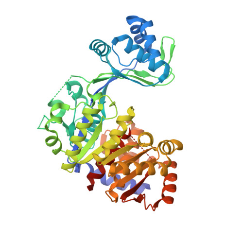
    Although ATP is the most common phosphoryl group donor for kinases, some kinases from certain hyperthermophilic archaea such as Pyrococcus horikoshii and Thermococcus litoralis use ADP as the phosphoryl donor. Those are ADP-dependent glucokinases (ADPGK) and phosphofructokinases in their glycolytic pathway. Here, we succeeded in gene cloning the ADPGK from P. horikoshii OT3 (phGK) in Escherichia coli,and in easy preparation of the enzyme, crystallization, and the structure determination of the apo enzyme. Recently, the three-dimensional structure of the ADPGK from T. litoralis (tlGK) in a complex with ADP was reported. The overall structure of two homologous enzymes (56.7%) was basically similar: This means that they consisted of large alpha/beta-domains and small domains. However, a marked adjustment of the two domains, which is a 10-A translation and a 20 degrees rotation from the conserved GG sequence located at the center of the hinge, was observed between the apo-phGK and ADP-tlGK structures. The ADP-binding loop (430-439) was disordered in the apo form. It is suggested that a large conformational change takes place during the enzymatic reaction.


  • Organizational Affiliation

    Institute for Health Sciences, Tokushima Bunri University, Yamashiro-cho, Japan.


Macromolecules
Find similar proteins by:  (by identity cutoff)  |  3D Structure
Entity ID: 1
MoleculeChains Sequence LengthOrganismDetailsImage
ADP-dependent Glucokinase457Pyrococcus horikoshiiMutation(s): 0 
EC: 2.7.1.2
UniProt
Find proteins for O58328 (Pyrococcus horikoshii (strain ATCC 700860 / DSM 12428 / JCM 9974 / NBRC 100139 / OT-3))
Explore O58328 
Go to UniProtKB:  O58328
Entity Groups  
Sequence Clusters30% Identity50% Identity70% Identity90% Identity95% Identity100% Identity
UniProt GroupO58328
Sequence Annotations
Expand
  • Reference Sequence
Experimental Data & Validation

Experimental Data

  • Method: X-RAY DIFFRACTION
  • Resolution: 2.00 Å
  • R-Value Free: 0.277 
  • R-Value Work: 0.213 
  • R-Value Observed: 0.213 
  • Space Group: P 21 21 21
Unit Cell:
Length ( Å )Angle ( ˚ )
a = 64.76α = 90
b = 74.73β = 90
c = 99.23γ = 90
Software Package:
Software NamePurpose
MOSFLMdata reduction
SCALAdata scaling
SOLVEphasing
CNSrefinement
CCP4data scaling

Structure Validation

View Full Validation Report


Entry History 

Deposition Data

Revision History  (Full details and data files)

  • Version 1.0: 2002-12-30
    Type: Initial release
  • Version 1.1: 2008-04-28
    Changes: Version format compliance
  • Version 1.2: 2011-07-13
    Changes: Version format compliance
  • Version 1.3: 2024-03-13
    Changes: Data collection, Database references