1L0I

Crystal structure of butyryl-ACP I62M mutant


Experimental Data Snapshot

  • Method: X-RAY DIFFRACTION
  • Resolution: 1.20 Å
  • R-Value Free: 0.193 
  • R-Value Work: 0.160 

wwPDB Validation   3D Report Full Report


This is version 1.4 of the entry. See complete history


Literature

X-ray Crystallographic Studies on Butyryl-ACP Reveal Flexibility of the Structure around a Putative Acyl Chain Binding Site

Roujeinikova, A.Baldock, C.Simon, W.J.Gilroy, J.Baker, P.J.Stuitje, A.R.Rice, D.W.Slabas, A.R.Rafferty, J.B.

(2002) Structure 10: 825-835

  • DOI: https://doi.org/10.1016/s0969-2126(02)00775-x
  • Primary Citation of Related Structures:  
    1L0H, 1L0I

  • PubMed Abstract: 

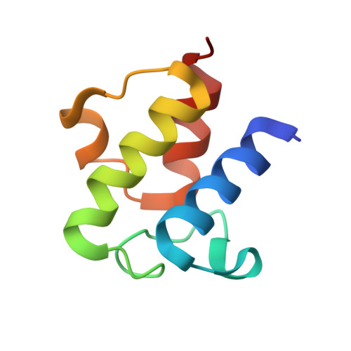
    Acyl carrier protein (ACP) is an essential cofactor in biosynthesis of fatty acids and many other reactions that require acyl transfer steps. We have determined the first crystal structures of an acylated form of ACP from E. coli, that of butyryl-ACP. Our analysis of the molecular surface of ACP reveals a plastic hydrophobic cavity in the vicinity of the phosphopantethylated Ser36 residue that is expanded and occupied by the butyryl and beta-mercaptoethylamine moieties of the acylated 4'-phosphopantetheine group in one of our crystal forms. In the other form, the cavity is contracted, and we propose that the protein has adopted the conformation after delivery of substrate into the active site of a partner enzyme.


  • Organizational Affiliation

    Krebs Institute for Biomolecular Research, Department of Molecular Biology and Biotechnology, The University of Sheffield, S10 2TN, Sheffield, United Kingdom.


Macromolecules
Find similar proteins by:  (by identity cutoff)  |  3D Structure
Entity ID: 1
MoleculeChains Sequence LengthOrganismDetailsImage
Acyl carrier protein78Escherichia coliMutation(s): 1 
UniProt
Find proteins for P0A6A8 (Escherichia coli (strain K12))
Explore P0A6A8 
Go to UniProtKB:  P0A6A8
Entity Groups  
Sequence Clusters30% Identity50% Identity70% Identity90% Identity95% Identity100% Identity
UniProt GroupP0A6A8
Sequence Annotations
Expand
  • Reference Sequence
Small Molecules
Ligands 4 Unique
IDChains Name / Formula / InChI Key2D Diagram3D Interactions
PSR
Query on PSR

Download Ideal Coordinates CCD File 
K [auth A]THIOBUTYRIC ACID S-{2-[3-(2-HYDROXY-3,3-DIMETHYL-4-PHOSPHONOOXY-BUTYRYLAMINO)-PROPIONYLAMINO]-ETHYL} ESTER
C15 H29 N2 O8 P S
PZIQDUSGQUZEBS-CYBMUJFWSA-N
CAC
Query on CAC

Download Ideal Coordinates CCD File 
D [auth A]CACODYLATE ION
C2 H6 As O2
OGGXGZAMXPVRFZ-UHFFFAOYSA-M
ZN
Query on ZN

Download Ideal Coordinates CCD File 
C [auth A]
E [auth A]
F [auth A]
G [auth A]
H [auth A]
C [auth A],
E [auth A],
F [auth A],
G [auth A],
H [auth A],
I [auth A],
J [auth A]
ZINC ION
Zn
PTFCDOFLOPIGGS-UHFFFAOYSA-N
NA
Query on NA

Download Ideal Coordinates CCD File 
B [auth A]SODIUM ION
Na
FKNQFGJONOIPTF-UHFFFAOYSA-N
Experimental Data & Validation

Experimental Data

  • Method: X-RAY DIFFRACTION
  • Resolution: 1.20 Å
  • R-Value Free: 0.193 
  • R-Value Work: 0.160 
  • Space Group: P 21 21 21
Unit Cell:
Length ( Å )Angle ( ˚ )
a = 27.347α = 90
b = 41.941β = 90
c = 64.533γ = 90
Software Package:
Software NamePurpose
Adxvdata processing
DENZOdata reduction
SCALEPACKdata scaling
warpntracemodel building
SHELXL-97refinement
WARPNTRACEphasing

Structure Validation

View Full Validation Report


Entry History 

Deposition Data

Revision History  (Full details and data files)

  • Version 1.0: 2003-02-11
    Type: Initial release
  • Version 1.1: 2008-04-28
    Changes: Version format compliance
  • Version 1.2: 2011-07-13
    Changes: Non-polymer description, Version format compliance
  • Version 1.3: 2017-10-11
    Changes: Advisory, Refinement description
  • Version 1.4: 2021-10-27
    Changes: Advisory, Database references, Derived calculations