1KWK

Crystal structure of Thermus thermophilus A4 beta-galactosidase in complex with galactose


Experimental Data Snapshot

  • Method: X-RAY DIFFRACTION
  • Resolution: 2.20 Å
  • R-Value Free: 0.200 
  • R-Value Work: 0.169 
  • R-Value Observed: 0.169 

wwPDB Validation   3D Report Full Report


Ligand Structure Quality Assessment 


This is version 1.4 of the entry. See complete history


Literature

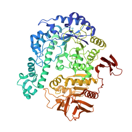
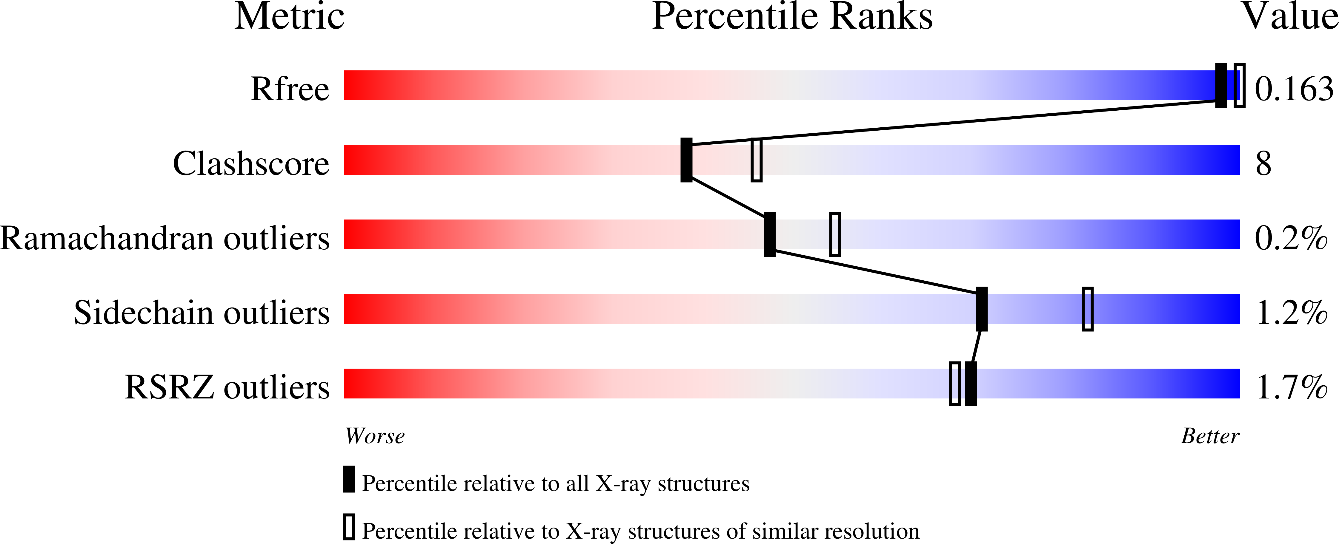
Trimeric crystal structure of the glycoside hydrolase family 42 beta-galactosidase from Thermus thermophilus A4 and the structure of its complex with galactose.

Hidaka, M.Fushinobu, S.Ohtsu, N.Motoshima, H.Matsuzawa, H.Shoun, H.Wakagi, T.

(2002) J Mol Biol 322: 79-91

  • DOI: https://doi.org/10.1016/s0022-2836(02)00746-5
  • Primary Citation of Related Structures:  
    1KWG, 1KWK

  • PubMed Abstract: 

    The beta-galactosidase from an extreme thermophile, Thermus thermophilus A4 (A4-beta-Gal), is thermostable and belongs to the glycoside hydrolase family 42 (GH-42). As the first known structures of a GH-42 enzyme, we determined the crystal structures of free and galactose-bound A4-beta-Gal at 1.6A and 2.2A resolution, respectively. A4-beta-Gal forms a homotrimeric structure resembling a flowerpot. Each monomer has an active site located inside a large central tunnel. The N-terminal domain of A4-beta-Gal has a TIM barrel fold, as predicted from hydrophobic cluster analysis. The putative catalytic residues of A4-beta-Gal (Glu141 and Glu312) superimpose well with the catalytic residues of Escherichia coli beta-galactosidase. The environment around the catalytic nucleophile (Glu312) is similar to that in the case of E.coli beta-galactosidase, but the recognition mechanism for a substrate is different. Trp182 of the next subunit of the trimer constitutes a part of the active-site pocket, indicating that the trimeric structure is essential for the enzyme activity. Structural comparison with other glycoside hydrolases revealed that many features of the 4/7 superfamily are conserved in the A4-beta-Gal structure. On the basis of the results of 1H NMR spectroscopy, A4-beta-Gal was determined to be a "retaining" enzyme. Interestingly, the active site was similar with those of retaining enzymes, but the overall fold of the TIM barrel domain was very similar to that of an inverting enzyme, beta-amylase.


  • Organizational Affiliation

    Department of Biotechnology, The University of Tokyo, Japan.


Macromolecules
Find similar proteins by:  (by identity cutoff)  |  3D Structure
Entity ID: 1
MoleculeChains Sequence LengthOrganismDetailsImage
BETA-GALACTOSIDASE645Thermus thermophilusMutation(s): 0 
EC: 3.2.1.23
UniProt
Find proteins for O69315 (Thermus thermophilus)
Explore O69315 
Go to UniProtKB:  O69315
Entity Groups  
Sequence Clusters30% Identity50% Identity70% Identity90% Identity95% Identity100% Identity
UniProt GroupO69315
Sequence Annotations
Expand
  • Reference Sequence
Small Molecules
Ligands 5 Unique
IDChains Name / Formula / InChI Key2D Diagram3D Interactions
GAL
Query on GAL

Download Ideal Coordinates CCD File 
B [auth A]beta-D-galactopyranose
C6 H12 O6
WQZGKKKJIJFFOK-FPRJBGLDSA-N
MPD
Query on MPD

Download Ideal Coordinates CCD File 
G [auth A],
H [auth A]
(4S)-2-METHYL-2,4-PENTANEDIOL
C6 H14 O2
SVTBMSDMJJWYQN-YFKPBYRVSA-N
ZN
Query on ZN

Download Ideal Coordinates CCD File 
F [auth A]ZINC ION
Zn
PTFCDOFLOPIGGS-UHFFFAOYSA-N
ACT
Query on ACT

Download Ideal Coordinates CCD File 
D [auth A],
E [auth A]
ACETATE ION
C2 H3 O2
QTBSBXVTEAMEQO-UHFFFAOYSA-M
CL
Query on CL

Download Ideal Coordinates CCD File 
C [auth A]CHLORIDE ION
Cl
VEXZGXHMUGYJMC-UHFFFAOYSA-M
Experimental Data & Validation

Experimental Data

  • Method: X-RAY DIFFRACTION
  • Resolution: 2.20 Å
  • R-Value Free: 0.200 
  • R-Value Work: 0.169 
  • R-Value Observed: 0.169 
  • Space Group: P 3 2 1
Unit Cell:
Length ( Å )Angle ( ˚ )
a = 97.592α = 90
b = 97.592β = 90
c = 128.898γ = 120
Software Package:
Software NamePurpose
MOSFLMdata reduction
SCALAdata scaling
SOLVEphasing
CNSrefinement
CCP4data scaling

Structure Validation

View Full Validation Report


Ligand Structure Quality Assessment 


Entry History 

Deposition Data

Revision History  (Full details and data files)

  • Version 1.0: 2002-10-02
    Type: Initial release
  • Version 1.1: 2008-04-27
    Changes: Version format compliance
  • Version 1.2: 2011-07-13
    Changes: Derived calculations, Version format compliance
  • Version 1.3: 2020-07-29
    Type: Remediation
    Reason: Carbohydrate remediation
    Changes: Advisory, Data collection, Derived calculations, Structure summary
  • Version 1.4: 2024-03-13
    Changes: Data collection, Database references, Structure summary