1JUX

1.6A Resolution Crystal Structures of the DNA Octamers d(IUATATAC) and d(ITITACAC):Binding of Two Distamycin Drugs Side-by-Side


Experimental Data Snapshot

  • Method: X-RAY DIFFRACTION
  • Resolution: 1.60 Å
  • R-Value Free: 0.179 
  • R-Value Work: 0.149 

wwPDB Validation   3D Report Full Report


Ligand Structure Quality Assessment 


This is version 1.4 of the entry. See complete history


Literature

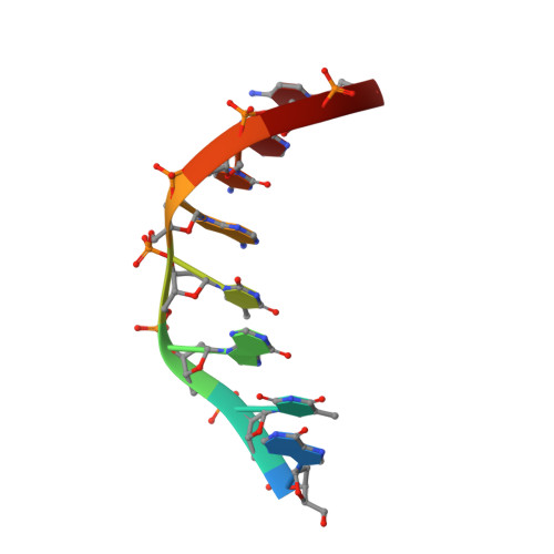
Structure of d(ITITACAC) complexed with distamycin at 1.6 A resolution.

Deng, J.Pan, B.Sundaralingam, M.

(2003) Acta Crystallogr D Biol Crystallogr 59: 2342-2344

  • DOI: https://doi.org/10.1107/s0907444903020730
  • Primary Citation of Related Structures:  
    1JUX

  • PubMed Abstract: 

    The crystal structure of the DNA octamer d(ITITACAC)(2) complexed with distamycin has been determined at 1.6 A resolution and refined to a final R(work) and R(free) of 17.0 and 20.7%, respectively. Two molecules of distamycin bind to the DNA duplex in an antiparallel side-by-side fashion. Each drug molecule covers five base pairs of the DNA duplex, with its amide groups hydrogen-bonding to bases in the proximal DNA strand. These two antiparallel drug molecules are stacked together with the pyrrole rings of one molecule stacking against the amide groups of the other. The present structure emphasizes the features of alternating DNA octamers in interaction with distamycin.


  • Organizational Affiliation

    Departments of Chemistry and Biochemistry, The Ohio State University, 200 Johnston Laboratory, 176 West 19th Avenue, Columbus, Ohio 43210, USA.


Macromolecules

Find similar nucleic acids by:  Sequence   |   3D Structure  

Entity ID: 1
MoleculeChains LengthOrganismImage
5'-D(*IP*TP*IP*TP*AP*CP*AP*C)-3'8N/A
Sequence Annotations
Expand
  • Reference Sequence
Small Molecules
Ligands 1 Unique
IDChains Name / Formula / InChI Key2D Diagram3D Interactions
DMY
Query on DMY

Download Ideal Coordinates CCD File 
B [auth A]DISTAMYCIN A
C22 H27 N9 O4
UPBAOYRENQEPJO-UHFFFAOYSA-N
Experimental Data & Validation

Experimental Data

  • Method: X-RAY DIFFRACTION
  • Resolution: 1.60 Å
  • R-Value Free: 0.179 
  • R-Value Work: 0.149 
  • Space Group: C 1 2 1
Unit Cell:
Length ( Å )Angle ( ˚ )
a = 33.26α = 90
b = 25.05β = 124.23
c = 28.3γ = 90
Software Package:
Software NamePurpose
AMoREphasing
CNSrefinement

Structure Validation

View Full Validation Report


Ligand Structure Quality Assessment 


Entry History 

Deposition Data

Revision History  (Full details and data files)

  • Version 1.0: 2004-01-27
    Type: Initial release
  • Version 1.1: 2008-04-27
    Changes: Version format compliance
  • Version 1.2: 2011-07-13
    Changes: Version format compliance
  • Version 1.3: 2017-10-04
    Changes: Refinement description
  • Version 1.4: 2024-02-07
    Changes: Data collection, Database references, Derived calculations