1JMM

Crystal structure of the V-region of Streptococcus mutans antigen I/II


Experimental Data Snapshot

  • Method: X-RAY DIFFRACTION
  • Resolution: 2.40 Å
  • R-Value Free: 0.234 
  • R-Value Work: 0.207 
  • R-Value Observed: 0.207 

wwPDB Validation   3D Report Full Report


This is version 1.2 of the entry. See complete history


Literature

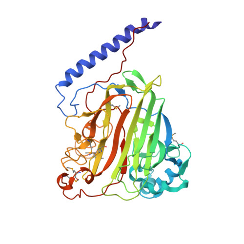
Crystal Structure of the V-region of Streptococcus mutans Antigen I/II at 2.4 a Resolution Suggests a Sugar Preformed Binding Site

Troffer-Charlier, N.Ogier, J.Moras, D.Cavarelli, J.

(2002) J Mol Biol 318: 179-188

  • DOI: https://doi.org/10.1016/S0022-2836(02)00025-6
  • Primary Citation of Related Structures:  
    1JMM

  • PubMed Abstract: 

    Antigens I/II are large multifunctional adhesins from oral viridans streptococci that exert immunomodulatory effects on human cells and play important roles in inflammatory disorders. Among them, Streptococcus mutans plays a major role in the initiation of dental caries. The structure of the V-region (SrV+, residues 464-840) of the antigen I/II of S. mutans has been determined using the multiwavelength anomalous diffraction phasing technique with seleno-methionine-substituted recombinant protein and subsequently refined at 2.4 A resolution. The crystal structure of SrV+ revealed a lectin-like fold that displays a putative preformed carbohydrate-binding site stabilized by a metal ion. Inhibition of this binding site may confer to humans a protection against dental caries and dissemination of the bacteria to extra-oral sites involved in life-threatening inflammatory diseases. This crystal structure constitutes a first step in understanding the structure-function relationship of antigens I/II and may help in delineating new preventive or therapeutic strategies against colonization of the host by oral streptococci.


  • Organizational Affiliation

    Institut National de la Santé et de la Recherche Médicale U424, Faculté de Chirurgie Dentaire, Université Louis Pasteur, 11 rue Humann, 67085 Strasbourg, France.


Macromolecules
Find similar proteins by:  (by identity cutoff)  |  3D Structure
Entity ID: 1
MoleculeChains Sequence LengthOrganismDetailsImage
protein I/II V-region377Streptococcus mutansMutation(s): 0 
UniProt
Find proteins for P11657 (Streptococcus mutans)
Explore P11657 
Go to UniProtKB:  P11657
Entity Groups  
Sequence Clusters30% Identity50% Identity70% Identity90% Identity95% Identity100% Identity
UniProt GroupP11657
Sequence Annotations
Expand
  • Reference Sequence
Small Molecules
Ligands 1 Unique
IDChains Name / Formula / InChI Key2D Diagram3D Interactions
NA
Query on NA

Download Ideal Coordinates CCD File 
B [auth A]SODIUM ION
Na
FKNQFGJONOIPTF-UHFFFAOYSA-N
Modified Residues  1 Unique
IDChains TypeFormula2D DiagramParent
MSE
Query on MSE
A
L-PEPTIDE LINKINGC5 H11 N O2 SeMET
Experimental Data & Validation

Experimental Data

  • Method: X-RAY DIFFRACTION
  • Resolution: 2.40 Å
  • R-Value Free: 0.234 
  • R-Value Work: 0.207 
  • R-Value Observed: 0.207 
  • Space Group: P 63 2 2
Unit Cell:
Length ( Å )Angle ( ˚ )
a = 124.57α = 90
b = 124.57β = 90
c = 147.692γ = 120
Software Package:
Software NamePurpose
DENZOdata reduction
SCALEPACKdata scaling
SOLVEphasing
CNSrefinement

Structure Validation

View Full Validation Report


Entry History 

Deposition Data

Revision History  (Full details and data files)

  • Version 1.0: 2002-07-17
    Type: Initial release
  • Version 1.1: 2008-04-27
    Changes: Version format compliance
  • Version 1.2: 2011-07-13
    Changes: Version format compliance