1JEK

Visna TM CORE STRUCTURE


Experimental Data Snapshot

  • Method: X-RAY DIFFRACTION
  • Resolution: 1.50 Å
  • R-Value Free: 0.265 
  • R-Value Work: 0.213 
  • R-Value Observed: 0.213 

wwPDB Validation   3D Report Full Report


This is version 1.4 of the entry. See complete history


Literature

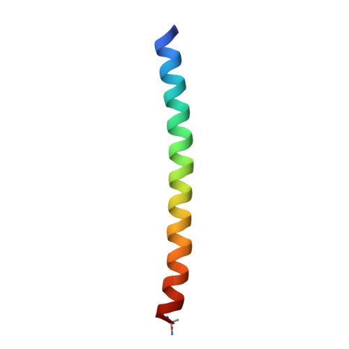
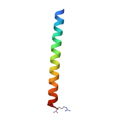
The trimer-of-hairpins motif in membrane fusion: Visna virus.

Malashkevich, V.N.Singh, M.Kim, P.S.

(2001) Proc Natl Acad Sci U S A 98: 8502-8506

  • DOI: https://doi.org/10.1073/pnas.151254798
  • Primary Citation of Related Structures:  
    1JEK

  • PubMed Abstract: 

    Structural studies of viral membrane fusion proteins suggest that a "trimer-of-hairpins" motif plays a critical role in the membrane fusion process of many enveloped viruses. In this motif, a coiled coil (formed by homotrimeric association of the N-terminal regions of the protein) is surrounded by three C-terminal regions that pack against the coiled coil in an oblique antiparallel manner. The resulting trimer-of-hairpins structure serves to bring the viral and cellular membranes together for fusion. learncoil-vmf, a computational program developed to recognize coiled coil-like regions that form the trimer-of-hairpins motif, predicts these regions in the membrane fusion protein of the Visna virus. Peptides corresponding to the computationally identified sequences were synthesized, and the soluble core of the Visna membrane fusion protein was reconstituted in solution. Its crystal structure at 1.5-A resolution demonstrates that a trimer-of-hairpins structure is formed. Remarkably, despite less than 23% sequence identity, the ectodomains in Visna and HIV-1 envelope glycoproteins show detailed structural conservation, especially within the area of a hydrophobic pocket in the central coiled coil currently being targeted for the development of new anti-HIV drugs.


  • Organizational Affiliation

    Howard Hughes Medical Institute, Whitehead Institute for Biomedical Research, Department of Biology, Massachusetts Institute of Technology, 9 Cambridge Center, Cambridge, MA 02142-1401, USA.


Macromolecules
Find similar proteins by:  (by identity cutoff)  |  3D Structure
Entity ID: 1
MoleculeChains Sequence LengthOrganismDetailsImage
ENV POLYPROTEIN42N/AMutation(s): 0 
UniProt
Find proteins for P35954 (Maedi visna virus (strain KV1772))
Explore P35954 
Go to UniProtKB:  P35954
Entity Groups  
Sequence Clusters30% Identity50% Identity70% Identity90% Identity95% Identity100% Identity
UniProt GroupP35954
Sequence Annotations
Expand
  • Reference Sequence
Find similar proteins by:  (by identity cutoff)  |  3D Structure
Entity ID: 2
MoleculeChains Sequence LengthOrganismDetailsImage
ENV POLYPROTEIN36N/AMutation(s): 0 
UniProt
Find proteins for P35954 (Maedi visna virus (strain KV1772))
Explore P35954 
Go to UniProtKB:  P35954
Entity Groups  
Sequence Clusters30% Identity50% Identity70% Identity90% Identity95% Identity100% Identity
UniProt GroupP35954
Sequence Annotations
Expand
  • Reference Sequence
Experimental Data & Validation

Experimental Data

  • Method: X-RAY DIFFRACTION
  • Resolution: 1.50 Å
  • R-Value Free: 0.265 
  • R-Value Work: 0.213 
  • R-Value Observed: 0.213 
  • Space Group: P 3 2 1
Unit Cell:
Length ( Å )Angle ( ˚ )
a = 52.548α = 90
b = 52.548β = 90
c = 61.686γ = 120
Software Package:
Software NamePurpose
AMoREphasing
CNSrefinement
DENZOdata reduction
SCALEPACKdata scaling

Structure Validation

View Full Validation Report


Entry History 

Deposition Data

Revision History  (Full details and data files)

  • Version 1.0: 2001-07-25
    Type: Initial release
  • Version 1.1: 2008-04-27
    Changes: Version format compliance
  • Version 1.2: 2011-07-13
    Changes: Derived calculations, Version format compliance
  • Version 1.3: 2011-11-16
    Changes: Atomic model
  • Version 1.4: 2023-08-16
    Changes: Data collection, Database references, Derived calculations, Refinement description