1J24

Crystal structure of archaeal XPF/Mus81 homolog, Hef from Pyrococcus furiosus, nuclease domain, Ca cocrystal


Experimental Data Snapshot

  • Method: X-RAY DIFFRACTION
  • Resolution: 1.78 Å
  • R-Value Free: 0.246 
  • R-Value Work: 0.215 

wwPDB Validation   3D Report Full Report


This is version 1.5 of the entry. See complete history


Literature

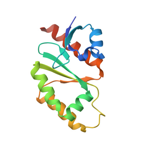
X-Ray and Biochemical Anatomy of an Archaeal XPF/Rad1/Mus81 Family Nuclease. Similarity between Its Endonuclease Domain and Restriction Enzymes

Nishino, T.Komori, K.Ishino, Y.Morikawa, K.

(2003) Structure 11: 445-457

  • DOI: https://doi.org/10.1016/s0969-2126(03)00046-7
  • Primary Citation of Related Structures:  
    1J22, 1J23, 1J24, 1J25

  • PubMed Abstract: 

    The XPF/Rad1/Mus81-dependent nuclease family specifically cleaves branched structures generated during DNA repair, replication, and recombination, and is essential for maintaining genome stability. Here, we report the domain organization of an archaeal homolog (Hef) of this family and the X-ray crystal structure of the middle domain, with the nuclease activity. The nuclease domain architecture exhibits remarkable similarity to those of restriction endonucleases, including the correspondence of the GDX(n)ERKX(3)D signature motif in Hef to the PDX(n)(E/D)XK motif in restriction enzymes. This structural study also suggests that the XPF/Rad1/Mus81/ERCC1 proteins form a dimer through each interface of the nuclease domain and the helix-hairpin-helix domain. Simultaneous disruptions of both interfaces result in their dissociation into separate monomers, with strikingly reduced endonuclease activities.


  • Organizational Affiliation

    Department of Structural Biology, Biomolecular Engineering Research Institute (BERI), 6-2-3 Furuedai, Suita, 565-0874, Osaka, Japan.


Macromolecules
Find similar proteins by:  (by identity cutoff)  |  3D Structure
Entity ID: 1
MoleculeChains Sequence LengthOrganismDetailsImage
ATP-dependent RNA helicase, putative143Pyrococcus furiosus DSM 3638Mutation(s): 2 
UniProt
Find proteins for Q8TZH8 (Pyrococcus furiosus (strain ATCC 43587 / DSM 3638 / JCM 8422 / Vc1))
Explore Q8TZH8 
Go to UniProtKB:  Q8TZH8
Entity Groups  
Sequence Clusters30% Identity50% Identity70% Identity90% Identity95% Identity100% Identity
UniProt GroupQ8TZH8
Sequence Annotations
Expand
  • Reference Sequence
Small Molecules
Ligands 1 Unique
IDChains Name / Formula / InChI Key2D Diagram3D Interactions
CA
Query on CA

Download Ideal Coordinates CCD File 
B [auth A]CALCIUM ION
Ca
BHPQYMZQTOCNFJ-UHFFFAOYSA-N
Experimental Data & Validation

Experimental Data

  • Method: X-RAY DIFFRACTION
  • Resolution: 1.78 Å
  • R-Value Free: 0.246 
  • R-Value Work: 0.215 
  • Space Group: P 31 2 1
Unit Cell:
Length ( Å )Angle ( ˚ )
a = 69.795α = 90
b = 69.795β = 90
c = 58.509γ = 120
Software Package:
Software NamePurpose
MOSFLMdata reduction
TRUNCATEdata reduction
SOLVEphasing
CNSrefinement
CCP4data scaling

Structure Validation

View Full Validation Report


Entry History 

Deposition Data

Revision History  (Full details and data files)

  • Version 1.0: 2003-04-22
    Type: Initial release
  • Version 1.1: 2008-04-27
    Changes: Version format compliance
  • Version 1.2: 2011-07-13
    Changes: Source and taxonomy, Version format compliance
  • Version 1.3: 2021-11-10
    Changes: Database references, Derived calculations
  • Version 1.4: 2023-12-27
    Changes: Data collection
  • Version 1.5: 2024-04-03
    Changes: Refinement description