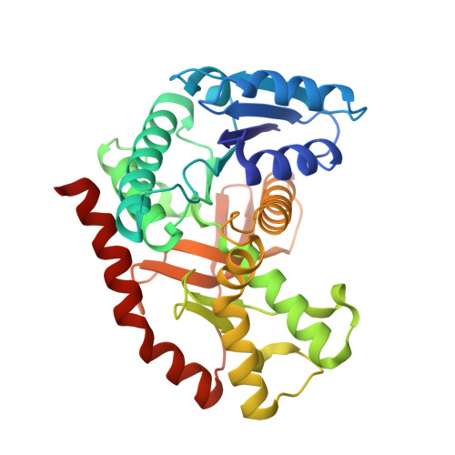
Structural Analyses of a Malate Dehydrogenase with a Variable Active Site
Bell, J.K., Yennawar, H.P., Wright, S.K., Thompson, J.R., Viola, R.E., Banaszak, L.J.(2001) J Biol Chem 276: 31156-31162
- PubMed: 11389141
- DOI: https://doi.org/10.1074/jbc.M100902200
- Primary Citation of Related Structures:
1IB6, 1IE3 - PubMed Abstract:
Malate dehydrogenase specifically oxidizes malate to oxaloacetate. The specificity arises from three arginines in the active site pocket that coordinate the carboxyl groups of the substrate and stabilize the newly forming hydroxyl/keto group during catalysis. Here, the role of Arg-153 in distinguishing substrate specificity is examined by the mutant R153C. The x-ray structure of the NAD binary complex at 2.1 A reveals two sulfate ions bound in the closed form of the active site. The sulfate that occupies the substrate binding site has been translated approximately 2 A toward the opening of the active site cavity. Its new location suggests that the low catalytic turnover observed in the R153C mutant may be due to misalignment of the hydroxyl or ketone group of the substrate with the appropriate catalytic residues. In the NAD.pyruvate ternary complex, the monocarboxylic inhibitor is bound in the open conformation of the active site. The pyruvate is coordinated not by the active site arginines, but through weak hydrogen bonds to the amide backbone. Energy minimized molecular models of unnatural analogues of R153C (Wright, S. K., and Viola, R. E. (2001) J. Biol. Chem. 276, 31151-31155) reveal that the regenerated amino and amido side chains can form favorable hydrogen-bonding interactions with the substrate, although a return to native enzymatic activity is not observed. The low activity of the modified R153C enzymes suggests that precise positioning of the guanidino side chain is essential for optimal orientation of the substrate.
Organizational Affiliation:
Department of Biochemistry, Molecular Biology and Biophysics, University of Minnesota, Minneapolis 55455, USA.