1ICI

CRYSTAL STRUCTURE OF A SIR2 HOMOLOG-NAD COMPLEX


Experimental Data Snapshot

  • Method: X-RAY DIFFRACTION
  • Resolution: 2.10 Å
  • R-Value Free: 0.249 
  • R-Value Work: 0.199 
  • R-Value Observed: 0.203 

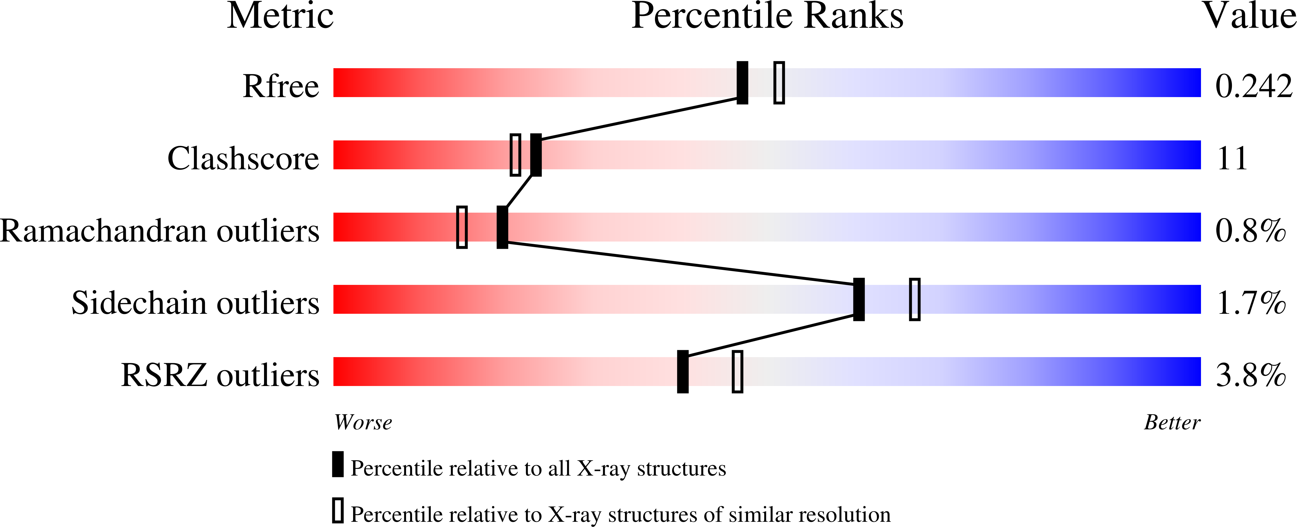
wwPDB Validation   3D Report Full Report


Ligand Structure Quality Assessment 


This is version 1.4 of the entry. See complete history


Literature

Crystal structure of a SIR2 homolog-NAD complex.

Min, J.Landry, J.Sternglanz, R.Xu, R.M.

(2001) Cell 105: 269-279

  • DOI: https://doi.org/10.1016/s0092-8674(01)00317-8
  • Primary Citation of Related Structures:  
    1ICI

  • PubMed Abstract: 

    The SIR2 protein family comprises a novel class of nicotinamide-adenine dinucleotide (NAD)-dependent protein deacetylases that function in transcriptional silencing, DNA repair, and life-span extension in Saccharomyces cerevisiae. Two crystal structures of a SIR2 homolog from Archaeoglobus fulgidus complexed with NAD have been determined at 2.1 A and 2.4 A resolutions. The structures reveal that the protein consists of a large domain having a Rossmann fold and a small domain containing a three-stranded zinc ribbon motif. NAD is bound in a pocket between the two domains. A distinct mode of NAD binding and an unusual configuration of the zinc ribbon motif are observed. The structures also provide important insights into the catalytic mechanism of NAD-dependent protein deacetylation by this family of enzymes.


  • Organizational Affiliation

    W. M. Keck Structural Biology Laboratory, Cold Spring Harbor Laboratory, Cold Spring Harbor, NY 11724, USA.


Macromolecules
Find similar proteins by:  (by identity cutoff)  |  3D Structure
Entity ID: 1
MoleculeChains Sequence LengthOrganismDetailsImage
TRANSCRIPTIONAL REGULATORY PROTEIN, SIR2 FAMILY
A, B
256Archaeoglobus fulgidusMutation(s): 0 
Gene Names: AF1676
UniProt
Find proteins for O28597 (Archaeoglobus fulgidus (strain ATCC 49558 / DSM 4304 / JCM 9628 / NBRC 100126 / VC-16))
Explore O28597 
Go to UniProtKB:  O28597
Entity Groups  
Sequence Clusters30% Identity50% Identity70% Identity90% Identity95% Identity100% Identity
UniProt GroupO28597
Sequence Annotations
Expand
  • Reference Sequence
Experimental Data & Validation

Experimental Data

  • Method: X-RAY DIFFRACTION
  • Resolution: 2.10 Å
  • R-Value Free: 0.249 
  • R-Value Work: 0.199 
  • R-Value Observed: 0.203 
  • Space Group: C 1 2 1
Unit Cell:
Length ( Å )Angle ( ˚ )
a = 65.41α = 90
b = 94.54β = 95.29
c = 93.78γ = 90
Software Package:
Software NamePurpose
PHASESphasing
CNSrefinement
MADNESSdata reduction
SCALEPACKdata scaling

Structure Validation

View Full Validation Report


Ligand Structure Quality Assessment 


Entry History 

Deposition Data

Revision History  (Full details and data files)

  • Version 1.0: 2001-05-02
    Type: Initial release
  • Version 1.1: 2008-04-27
    Changes: Version format compliance
  • Version 1.2: 2011-07-13
    Changes: Derived calculations, Version format compliance
  • Version 1.3: 2017-10-04
    Changes: Refinement description
  • Version 1.4: 2024-02-07
    Changes: Data collection, Database references, Derived calculations