1IB4

Crystal Structure of Polygalacturonase from Aspergillus Aculeatus at Ph4.5


Experimental Data Snapshot

  • Method: X-RAY DIFFRACTION
  • Resolution: 2.00 Å
  • R-Value Free: 0.217 
  • R-Value Work: 0.168 
  • R-Value Observed: 0.168 

wwPDB Validation   3D Report Full Report


Ligand Structure Quality Assessment 


This is version 2.0 of the entry. See complete history


Literature

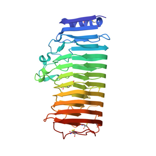
The X-ray structure of Aspergillus aculeatus polygalacturonase and a modeled structure of the polygalacturonase-octagalacturonate complex.

Cho, S.W.Lee, S.Shin, W.

(2001) J Mol Biol 311: 863-878

  • DOI: https://doi.org/10.1006/jmbi.2001.4919
  • Primary Citation of Related Structures:  
    1IA5, 1IB4

  • PubMed Abstract: 

    Polygalacturonases hydrolyze the alpha-(1-4) glycosidic bonds of de-esterified pectate in the smooth region of the plant cell wall. Crystal structures of polygalacturonase from Aspergillus aculeatus were determined at pH 4.5 and 8.5 both to 2.0 A resolution. A. aculeatus polygalacturonase is a glycoprotein with one N and ten O-glycosylation sites and folds into a right-handed parallel beta-helix. The structures of the three independent molecules are essentially the same, showing no dependency on pH or crystal packing, and are very similar to that of Aspergillus niger polygalacturonase. However, the structures of the long T1 loop containing a catalytic tyrosine residue are significantly different in the two proteins. A three-dimensional model showing the substrate binding mode for a family 28 hydrolase was obtained by a combined approach of flexible docking, molecular dynamics simulations, and energy minimization. The octagalacturonate substrate was modeled as an unbent irregular helix with the -1 ring in a half-chair ((4)H(3)) form that approaches the transition state conformation. A comparative modeling of the three polygalacturonases with known structure shows that six subsites ranging from -4 to +2 are clearly defined but subsites -5 and +3 may or may not be shaped depending on the nearby amino acid residues. Both distal subsites are mostly exposed to the solvent region and have weak binding affinity even if they exist. The complex model provides a clear explanation for the functions, either in catalysis or in substrate binding, of all conserved amino acid residues in the polygalacturonase family of proteins. Modeling suggests that the role of the conserved Asn157 and Tyr270, which had previously been unidentified, may be in transition state stabilization. In A. niger polygalacturonase, the long T1 loop may have to undergo conformational change upon binding of the substrate to bring the tyrosine residue close to subsite -1.


  • Organizational Affiliation

    School of Chemistry and Molecular Engineering, and Center for Molecular Catalysis, Seoul National University, Seoul, 151-742, Korea.


Macromolecules
Find similar proteins by:  (by identity cutoff)  |  3D Structure
Entity ID: 1
MoleculeChains Sequence LengthOrganismDetailsImage
POLYGALACTURONASE
A, B
339Aspergillus aculeatusMutation(s): 0 
EC: 3.2.1.15
UniProt
Find proteins for O74213 (Aspergillus aculeatus)
Explore O74213 
Go to UniProtKB:  O74213
Entity Groups  
Sequence Clusters30% Identity50% Identity70% Identity90% Identity95% Identity100% Identity
UniProt GroupO74213
Sequence Annotations
Expand
  • Reference Sequence
Oligosaccharides

Help

Entity ID: 2
MoleculeChains Length2D Diagram Glycosylation3D Interactions
alpha-D-mannopyranose-(1-4)-2-acetamido-2-deoxy-beta-D-glucopyranose-(1-4)-2-acetamido-2-deoxy-beta-D-glucopyranose
C, D
3N-Glycosylation
Glycosylation Resources
GlyTouCan:  G62182OO
GlyCosmos:  G62182OO
GlyGen:  G62182OO
Small Molecules
Ligands 2 Unique
IDChains Name / Formula / InChI Key2D Diagram3D Interactions
MAN
Query on MAN

Download Ideal Coordinates CCD File 
E [auth A]
F [auth A]
G [auth A]
H [auth A]
I [auth A]
E [auth A],
F [auth A],
G [auth A],
H [auth A],
I [auth A],
J [auth A],
K [auth A],
L [auth A],
M [auth A],
N [auth A],
O [auth B],
P [auth B],
Q [auth B],
R [auth B],
S [auth B],
T [auth B],
U [auth B],
V [auth B],
W [auth B],
X [auth B]
alpha-D-mannopyranose
C6 H12 O6
WQZGKKKJIJFFOK-PQMKYFCFSA-N
CD
Query on CD

Download Ideal Coordinates CCD File 
AA [auth B],
Y [auth B],
Z [auth B]
CADMIUM ION
Cd
WLZRMCYVCSSEQC-UHFFFAOYSA-N
Experimental Data & Validation

Experimental Data

  • Method: X-RAY DIFFRACTION
  • Resolution: 2.00 Å
  • R-Value Free: 0.217 
  • R-Value Work: 0.168 
  • R-Value Observed: 0.168 
  • Space Group: P 1 21 1
Unit Cell:
Length ( Å )Angle ( ˚ )
a = 56.1α = 90
b = 96.4β = 107.3
c = 57.77γ = 90
Software Package:
Software NamePurpose
MADNESSdata collection
SCALAdata scaling
CNSrefinement
SHELXL-97refinement
MADNESSdata reduction
CCP4data scaling
CNSphasing

Structure Validation

View Full Validation Report


Ligand Structure Quality Assessment 


Entry History 

Deposition Data

Revision History  (Full details and data files)

  • Version 1.0: 2001-09-19
    Type: Initial release
  • Version 1.1: 2008-04-27
    Changes: Version format compliance
  • Version 1.2: 2011-07-13
    Changes: Non-polymer description, Version format compliance
  • Version 1.3: 2014-12-17
    Changes: Derived calculations
  • Version 1.4: 2017-10-04
    Changes: Refinement description
  • Version 2.0: 2020-07-29
    Type: Remediation
    Reason: Carbohydrate remediation
    Changes: Atomic model, Data collection, Derived calculations, Structure summary