1HLC

X-RAY CRYSTAL STRUCTURE OF THE HUMAN DIMERIC S-LAC LECTIN, L-14-II, IN COMPLEX WITH LACTOSE AT 2.9 ANGSTROMS RESOLUTION


Experimental Data Snapshot

  • Method: X-RAY DIFFRACTION
  • Resolution: 2.90 Å
  • R-Value Work: 0.177 
  • R-Value Observed: 0.177 

wwPDB Validation   3D Report Full Report


This is version 2.1 of the entry. See complete history


Literature

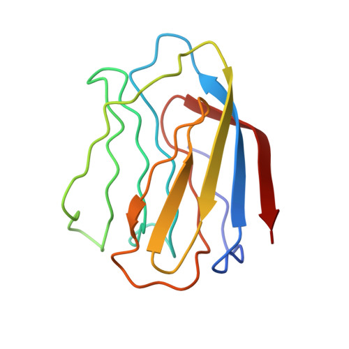
X-ray crystal structure of the human dimeric S-Lac lectin, L-14-II, in complex with lactose at 2.9-A resolution.

Lobsanov, Y.D.Gitt, M.A.Leffler, H.Barondes, S.H.Rini, J.M.

(1993) J Biol Chem 268: 27034-27038

  • DOI: https://doi.org/10.2210/pdb1hlc/pdb
  • Primary Citation of Related Structures:  
    1HLC

  • PubMed Abstract: 

    S-Lac lectins are a family of soluble lactose-binding animal lectins, some of which have been implicated in modulating cell-cell and cell-matrix interactions through specific carbohydrate-mediated recognition. We report here the x-ray crystal structure of a representative member of this family, the human dimeric S-Lac lectin, L-14-II, in complex with lactose, at 2.9-A resolution. The two-fold symmetric dimer is made up of two extended anti-parallel beta-sheets, which associate in a beta-sandwich motif. Remarkably, the L-14-II monomer shares not only the same topology, but a very similar beta-sheet structure with that of the leguminous plant lectins, suggesting a conserved structure-function relationship. Carbohydrate binding by L-14-II was found to involve protein residues that are very highly conserved among all S-Lac lectins. These residues map to a single DNA exon, suggesting a carbohydrate binding cassette common to all S-Lac lectins.


  • Organizational Affiliation

    Department of Molecular and Medical Genetics, University of Toronto, Ontario, Canada.


Macromolecules
Find similar proteins by:  (by identity cutoff)  |  3D Structure
Entity ID: 1
MoleculeChains Sequence LengthOrganismDetailsImage
HUMAN LECTIN
A, B
129Homo sapiensMutation(s): 0 
Gene Names: CDNA
UniProt & NIH Common Fund Data Resources
Find proteins for P05162 (Homo sapiens)
Explore P05162 
Go to UniProtKB:  P05162
PHAROS:  P05162
GTEx:  ENSG00000100079 
Entity Groups  
Sequence Clusters30% Identity50% Identity70% Identity90% Identity95% Identity100% Identity
UniProt GroupP05162
Sequence Annotations
Expand
  • Reference Sequence
Oligosaccharides

Help

Entity ID: 2
MoleculeChains Length2D Diagram Glycosylation3D Interactions
beta-D-galactopyranose-(1-4)-beta-D-glucopyranose
C, D
2N/A
Glycosylation Resources
GlyTouCan:  G84224TW
GlyCosmos:  G84224TW
GlyGen:  G84224TW
Biologically Interesting Molecules (External Reference) 1 Unique
Experimental Data & Validation

Experimental Data

  • Method: X-RAY DIFFRACTION
  • Resolution: 2.90 Å
  • R-Value Work: 0.177 
  • R-Value Observed: 0.177 
  • Space Group: P 21 21 21
Unit Cell:
Length ( Å )Angle ( ˚ )
a = 43.6α = 90
b = 57.8β = 90
c = 108.2γ = 90
Software Package:
Software NamePurpose
X-PLORmodel building
X-PLORrefinement
X-PLORphasing

Structure Validation

View Full Validation Report


Entry History 

Deposition Data

Revision History  (Full details and data files)

  • Version 1.0: 1994-04-30
    Type: Initial release
  • Version 1.1: 2008-03-24
    Changes: Version format compliance
  • Version 1.2: 2011-07-13
    Changes: Version format compliance
  • Version 2.0: 2020-07-29
    Type: Remediation
    Reason: Carbohydrate remediation
    Changes: Atomic model, Data collection, Derived calculations, Other, Structure summary
  • Version 2.1: 2024-02-07
    Changes: Data collection, Database references, Structure summary