Mimicking Natural Evolution in Vitro: An N-Acetylneuraminate Lyase Mutant with an Increased Dihydrodipicolinate Synthase Activity
Joerger, A.C., Mayer, S., Fersht, A.R.(2003) Proc Natl Acad Sci U S A 100: 5694
- PubMed: 12711733
- DOI: https://doi.org/10.1073/pnas.0531477100
- Primary Citation of Related Structures:
1HL2 - PubMed Abstract:
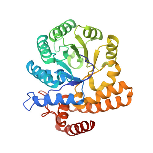
N-acetylneuraminate lyase (NAL) and dihydrodipicolinate synthase (DHDPS) belong to the NAL subfamily of (betaalpha)(8)-barrels. They share a common catalytic step but catalyze reactions in different biological pathways. By rational design, we have introduced various mutations into the NAL scaffold from Escherichia coli to switch the activity toward DHDPS. These mutants were tested with respect to their catalytic properties in vivo and in vitro as well as their stability. One point mutation (L142R) was sufficient to create an enzyme that could complement a bacterial auxotroph lacking the gene for DHDPS as efficiently as DHDPS itself. In vitro, this mutant had an increased DHDPS activity of up to 19-fold as defined by the specificity constant k(cat)K(M) for the new substrate l-aspartate-beta-semialdehyde when compared with the residual activity of NAL wild-type, mainly because of an increased turnover rate. At the same time, mutant L142R maintained much of its original NAL activity. We have solved the crystal structure of mutant L142R at 1.8 A resolution in complex with the inhibitor beta-hydroxypyruvate. This structure reveals that the conformations of neighboring active site residues are left virtually unchanged by the mutation. The high flexibility of R142 may favor its role in assisting in catalysis. Perhaps, nature has exploited the catalytic promiscuity of many enzymes to evolve novel enzymes or biological pathways during the course of evolution.
Organizational Affiliation:
Cambridge University Chemical Laboratory and Cambridge Centre for Protein Engineering, Medical Research Council Centre, Hills Road, Cambridge CB2 2QH, United Kingdom.