1HIA

KALLIKREIN COMPLEXED WITH HIRUSTASIN


Experimental Data Snapshot

  • Method: X-RAY DIFFRACTION
  • Resolution: 2.40 Å
  • R-Value Free: 0.311 
  • R-Value Work: 0.205 
  • R-Value Observed: 0.205 

wwPDB Validation   3D Report Full Report


This is version 1.3 of the entry. See complete history


Literature

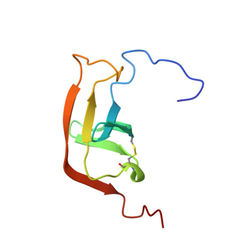
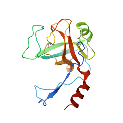
A new structural class of serine protease inhibitors revealed by the structure of the hirustasin-kallikrein complex.

Mittl, P.R.Di Marco, S.Fendrich, G.Pohlig, G.Heim, J.Sommerhoff, C.Fritz, H.Priestle, J.P.Grutter, M.G.

(1997) Structure 5: 253-264

  • DOI: https://doi.org/10.1016/s0969-2126(97)00183-4
  • Primary Citation of Related Structures:  
    1HIA

  • PubMed Abstract: 

    Hirustasin belongs to a class of serine protease inhibitors characterized by a well conserved pattern of cysteine residues. Unlike the closely related inhibitors, antistasin/ghilanten and guamerin, which are selective for coagulation factor Xa or neutrophil elastase, hirustasin binds specifically to tissue kallikrein. The conservation of the pattern of cysteine residues and the significant sequence homology suggest that these related inhibitors possess a similar three-dimensional structure to hirustasin. The crystal structure of the complex between tissue kallikrein and hirustasin was analyzed at 2.4 resolution. Hirustasin folds into a brick-like structure that is dominated by five disulfide bridges and is sparse in secondary structural elements. The cysteine residues are connected in an abab cdecde pattern that causes the polypeptide chain to fold into two similar motifs. As a hydrophobic core is absent from hirustasin the disulfide bridges maintain the tertiary structure and present the primary binding loop to the active site of the protease. The general structural topography and disulfide connectivity of hirustasin has not previously been described. The crystal structure of the kallikrein-hirustasin complex reveals that hirustasin differs from other serine protease inhibitors in its conformation and its disulfide bond connectivity, making it the prototype for a new class of inhibitor. The disulfide pattern shows that the structure consists of two domains, but only the C-terminal domain interacts with the protease. The disulfide pattern of the N-terminal domain is related to the pattern found in other proteins. Kallikrein recognizes hirustasin by the formation of an antiparallel beta sheet between the protease and the inhibitor. The P1 arginine binds in a deep negatively charged pocket of the enzyme. An additional pocket at the periphery of the active site accommodates the sidechain of the P4 valine.


  • Organizational Affiliation

    CDDT Pharmaceutical Research, Ciba-Geigy Ltd., 4002 Basel, Switzerland.


Macromolecules
Find similar proteins by:  (by identity cutoff)  |  3D Structure
Entity ID: 1
MoleculeChains Sequence LengthOrganismDetailsImage
KALLIKREINA,
D [auth X]
80Sus scrofaMutation(s): 0 
EC: 3.4.21.35
UniProt
Find proteins for P00752 (Sus scrofa)
Explore P00752 
Go to UniProtKB:  P00752
Entity Groups  
Sequence Clusters30% Identity50% Identity70% Identity90% Identity95% Identity100% Identity
UniProt GroupP00752
Sequence Annotations
Expand
  • Reference Sequence
Find similar proteins by:  (by identity cutoff)  |  3D Structure
Entity ID: 2
MoleculeChains Sequence LengthOrganismDetailsImage
KALLIKREINB,
E [auth Y]
152Sus scrofaMutation(s): 0 
EC: 3.4.21.35
UniProt
Find proteins for P00752 (Sus scrofa)
Explore P00752 
Go to UniProtKB:  P00752
Entity Groups  
Sequence Clusters30% Identity50% Identity70% Identity90% Identity95% Identity100% Identity
UniProt GroupP00752
Sequence Annotations
Expand
  • Reference Sequence
Find similar proteins by:  (by identity cutoff)  |  3D Structure
Entity ID: 3
MoleculeChains Sequence LengthOrganismDetailsImage
HIRUSTASINC [auth I],
F [auth J]
48Hirudo medicinalisMutation(s): 0 
UniProt
Find proteins for P80302 (Hirudo medicinalis)
Explore P80302 
Go to UniProtKB:  P80302
Entity Groups  
Sequence Clusters30% Identity50% Identity70% Identity90% Identity95% Identity100% Identity
UniProt GroupP80302
Sequence Annotations
Expand
  • Reference Sequence
Experimental Data & Validation

Experimental Data

  • Method: X-RAY DIFFRACTION
  • Resolution: 2.40 Å
  • R-Value Free: 0.311 
  • R-Value Work: 0.205 
  • R-Value Observed: 0.205 
  • Space Group: P 21 21 2
Unit Cell:
Length ( Å )Angle ( ˚ )
a = 116.9α = 90
b = 86β = 90
c = 69.4γ = 90
Software Package:
Software NamePurpose
MARXDSdata collection
MARSCALEdata reduction
AMoREphasing
X-PLORrefinement
MARXDSdata reduction
MARSCALEdata scaling

Structure Validation

View Full Validation Report


Entry History 

Deposition Data

Revision History  (Full details and data files)

  • Version 1.0: 1997-12-24
    Type: Initial release
  • Version 1.1: 2008-03-24
    Changes: Version format compliance
  • Version 1.2: 2011-07-13
    Changes: Version format compliance
  • Version 1.3: 2023-08-09
    Changes: Database references, Refinement description