1HH7

REFINED CRYSTAL STRUCTURE OF CYTOCHROME C2 FROM RHODOPSEUDOMONAS PALUSTRIS AT 1.4 ANGSTROM RESOLUTION


Experimental Data Snapshot

  • Method: X-RAY DIFFRACTION
  • Resolution: 1.40 Å
  • R-Value Observed: 0.151 

wwPDB Validation   3D Report Full Report


This is version 2.1 of the entry. See complete history


Literature

Crystallization and Preliminary X-Ray Analysis of Two Ph-Dependent Forms of Cytochrome C2 from Rhodopseudomonas Palustris

Garau, G.Geremia, S.Randaccio, L.Vaccari, L.Viezzoli, M.S.

(2000) Acta Crystallogr D Biol Crystallogr 56: 1699

  • DOI: https://doi.org/10.1107/s0907444900013573
  • Primary Citation of Related Structures:  
    1HH7

  • PubMed Abstract: 

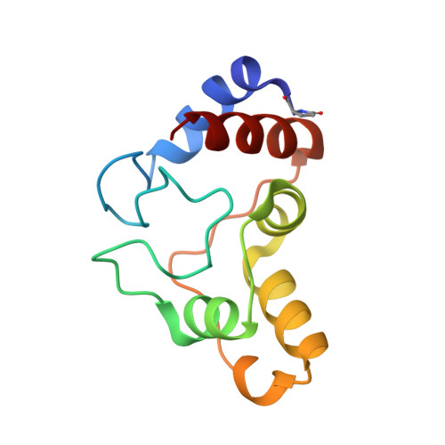
    Cytochrome c(2) from Rhodopseudomonas palustris has been crystallized in two different crystal forms: a monoclinic form I at pH 4.4 from both reduced and oxidized protein solution and a trigonal form II at pH 9.0 from reduced protein solution. Complete 1. 7 and 1.4 A resolution data sets were collected from the oxidized form I and from the form II, respectively. The preliminary structures show an important change in the iron coordination environment in the trigonal form obtained at basic pH arising from the substitution of the Met ligand by an ammonia molecule.


  • Organizational Affiliation

    Department of Chemical Sciences, University of Trieste, Via L. Giorgieri 1, I-34127 Trieste, Italy.


Macromolecules
Find similar proteins by:  (by identity cutoff)  |  3D Structure
Entity ID: 1
MoleculeChains Sequence LengthOrganismDetailsImage
CYTOCHROME C2114Rhodopseudomonas palustrisMutation(s): 0 
UniProt
Find proteins for P00091 (Rhodopseudomonas palustris (strain ATCC BAA-98 / CGA009))
Explore P00091 
Go to UniProtKB:  P00091
Entity Groups  
Sequence Clusters30% Identity50% Identity70% Identity90% Identity95% Identity100% Identity
UniProt GroupP00091
Sequence Annotations
Expand
  • Reference Sequence
Small Molecules
Modified Residues  1 Unique
IDChains TypeFormula2D DiagramParent
PCA
Query on PCA
A
L-PEPTIDE LINKINGC5 H7 N O3GLN
Experimental Data & Validation

Experimental Data

  • Method: X-RAY DIFFRACTION
  • Resolution: 1.40 Å
  • R-Value Observed: 0.151 
  • Space Group: P 32 2 1
Unit Cell:
Length ( Å )Angle ( ˚ )
a = 64.67α = 90
b = 64.67β = 90
c = 68.25γ = 120
Software Package:
Software NamePurpose
SHELXL-97refinement
MOSFLMdata reduction
SCALAdata scaling
AMoREphasing

Structure Validation

View Full Validation Report


Entry History 

Deposition Data

Revision History  (Full details and data files)

  • Version 1.0: 2001-01-02
    Type: Initial release
  • Version 1.1: 2011-05-08
    Changes: Version format compliance
  • Version 1.2: 2011-07-13
    Changes: Version format compliance
  • Version 1.3: 2019-05-22
    Changes: Data collection, Derived calculations, Other, Refinement description
  • Version 2.0: 2020-03-11
    Changes: Advisory, Polymer sequence
  • Version 2.1: 2023-12-13
    Changes: Data collection, Database references, Derived calculations, Refinement description