1HF2

Crystal structure of the bacterial cell-division inhibitor MinC from T. maritima


Experimental Data Snapshot

  • Method: X-RAY DIFFRACTION
  • Resolution: 2.20 Å
  • R-Value Free: 0.300 
  • R-Value Work: 0.237 
  • R-Value Observed: 0.237 

wwPDB Validation   3D Report Full Report


This is version 2.0 of the entry. See complete history


Literature

Crystal Structure of the Bacterial Cell-Division Inhibitor Minc

Cordell, S.C.Anderson, R.E.Lowe, J.

(2001) EMBO J 20: 2454

  • DOI: https://doi.org/10.1093/emboj/20.10.2454
  • Primary Citation of Related Structures:  
    1HF2

  • PubMed Abstract: 

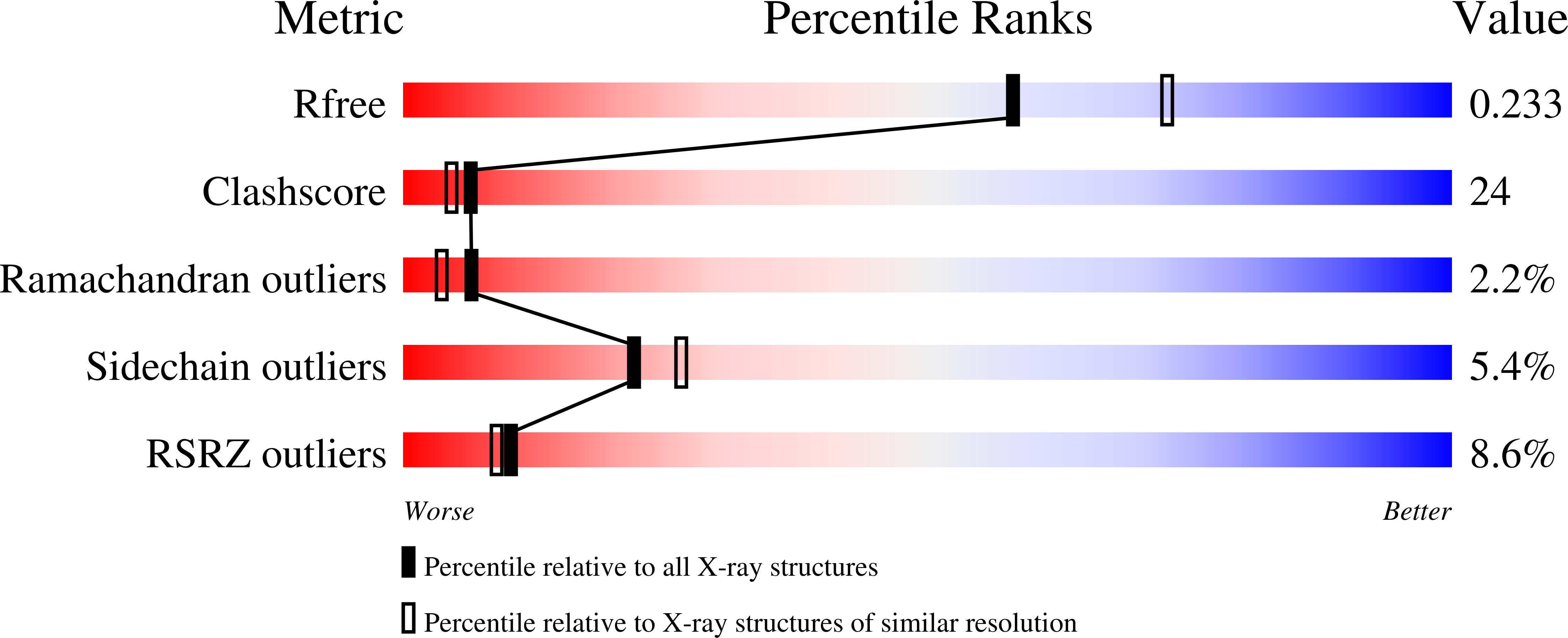
    Bacterial cell division requires accurate selection of the middle of the cell, where the bacterial tubulin homologue FtsZ polymerizes into a ring structure. In Escherichia coli, site selection is dependent on MinC, MinD and MINE: MinC acts, with MinD, to inhibit division at sites other than the midcell by directly interacting with FTSZ: Here we report the crystal structure to 2.2 A of MinC from Thermotoga maritima. MinC consists of two domains separated by a short linker. The C-terminal domain is a right-handed beta-helix and is involved in dimer formation. The crystals contain two different MinC dimers, demonstrating flexibility in the linker region. The two-domain architecture and dimerization of MinC can be rationalized with a model of cell division inhibition. MinC does not act like SulA, which affects the GTPase activity of FtsZ, and the model can explain how MinC would select for the FtsZ polymer rather than the monomer.


  • Organizational Affiliation

    MRC Laboratory of Molecular Biology, Hills Road, Cambridge CB2 2QH, UK.


Macromolecules
Find similar proteins by:  (by identity cutoff)  |  3D Structure
Entity ID: 1
MoleculeChains Sequence LengthOrganismDetailsImage
SEPTUM SITE-DETERMINING PROTEIN MINC
A, B, C, D
210Thermotoga maritimaMutation(s): 0 
Gene Names: MINC
UniProt
Find proteins for Q9X0D7 (Thermotoga maritima (strain ATCC 43589 / DSM 3109 / JCM 10099 / NBRC 100826 / MSB8))
Explore Q9X0D7 
Go to UniProtKB:  Q9X0D7
Entity Groups  
Sequence Clusters30% Identity50% Identity70% Identity90% Identity95% Identity100% Identity
UniProt GroupQ9X0D7
Sequence Annotations
Expand
  • Reference Sequence
Experimental Data & Validation

Experimental Data

  • Method: X-RAY DIFFRACTION
  • Resolution: 2.20 Å
  • R-Value Free: 0.300 
  • R-Value Work: 0.237 
  • R-Value Observed: 0.237 
  • Space Group: P 21 21 21
Unit Cell:
Length ( Å )Angle ( ˚ )
a = 51.58α = 90
b = 106.09β = 90
c = 162.53γ = 90
Software Package:
Software NamePurpose
CNSrefinement
MOSFLMdata reduction
SCALAdata scaling
SOLVEphasing

Structure Validation

View Full Validation Report


Entry History 

Deposition Data

Revision History  (Full details and data files)

  • Version 1.0: 2001-05-30
    Type: Initial release
  • Version 1.1: 2011-05-08
    Changes: Version format compliance
  • Version 1.2: 2011-07-13
    Changes: Version format compliance
  • Version 2.0: 2018-01-24
    Changes: Atomic model, Source and taxonomy