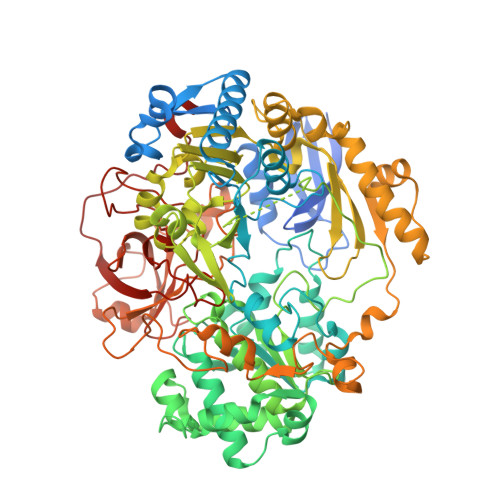
Reactions of Dimethyl Sulphoxide Reductase in the Presence of Dimethylsulphide and the Structure of the Dimethylsulphide-Modified Enzyme
Bray, R.C., Adams, B., Smith, A.T., Richards, R.L., Lowe, D.J., Bailey, S.(2001) Biochemistry 40: 9810
- PubMed: 11502174
- DOI: https://doi.org/10.1021/bi010559r
- Primary Citation of Related Structures:
1H5N - PubMed Abstract:
The bis-molybdopterin enzyme dimethylsulfoxide reductase (DMSOR) from Rhodobacter capsulatus catalyzes the conversion of dimethyl sulfoxide (DMSO) to dimethyl sulfide (DMS), reversibly, in the presence of suitable e(-)-donors or e(-)-acceptors. The catalytically significant intermediate formed by reaction of DMSOR with DMS ('the DMS species') and a damaged enzyme form derived by reaction of the latter with O(2) (DMS-modified enzyme, DMSOR(mod)D) have been investigated. Evidence is presented that Mo in the DMS species is not, as widely assumed, Mo(IV). Formation of the DMS species is reversed on removing DMS or by addition of an excess of DMSO. Equilibrium constants for the competing reactions of DMS and DMSO with the oxidized enzyme (K(d) = 0.07 +/- 0.01 and 21 +/- 5 mM, respectively) that control these processes indicate formation of the DMS species occurs at a redox potential that is 80 mV higher than that required, according to the literature, for reduction of Mo(VI) to Mo(IV) in the free enzyme. Specificity studies show that with dimethyl selenide, DMSOR yields a species analogous to the DMS species but with the 550 nm peak blue-shifted by 27 nm. It is concluded from published redox potential data that this band is due to metal-to-ligand charge transfer from Mo(V) to the chalcogenide. Since the DMS species gives no EPR signal in the normal or parallel mode, a free radical is presumed to be in close proximity to the metal, most likely on the S. The species is thus formulated as Mo(V)-O-S(*)Me(2). Existing X-ray crystallographic and Raman data are consistent with this structure. Furthermore, 1e(-) oxidation of the DMS species with phenazine ethosulfate yields a Mo(V) form without an -OH ligand, since its EPR signal shows no proton splittings. This form presumably arises via dissociation of DMSO. The structure of DMSOR(mod)D has been determined by X-ray crystallography. All four thiolate ligands and Ogamma of serine-147 remain coordinated to Mo, but there are no terminal oxygen ligands and Mo is Mo(VI). Thus, it is a dead-end species, neither oxo group acceptance nor e(-)-donation being possible. O(2)-dependent formation of DMSOR(mod)D represents noncatalytic breakdown of the DMS species by a pathway alternative to that in turnover, with oxidation to Mo(VI) presumably preceding product release. Steps in the forward and backward catalytic cycles are discussed in relation to earlier stopped-flow data. The finding that in the back-assay the Mo(IV) state may at least in part be by-passed via two successive 1e(-) reactions of the DMS species with the e(-)-acceptor, may have implications in relation to the existence of separate molybdopterin enzymes catalyzing DMSO reduction and DMS oxidation, respectively.
Organizational Affiliation:
School of Chemistry, Physics, and Environmental Science, University of Sussex, Brighton, BN1 9QJ, UK. r.c.bray@sussex.ac.uk