1GZ1

Mutant D416A of the CELLOBIOHYDROLASE CEL6A FROM HUMICOLA INSOLENS in complex with methyl-cellobiosyl-4-deoxy-4-thio-beta-D-cellobioside


Experimental Data Snapshot

  • Method: X-RAY DIFFRACTION
  • Resolution: 1.90 Å
  • R-Value Free: 0.211 
  • R-Value Work: 0.166 
  • R-Value Observed: 0.168 

wwPDB Validation   3D Report Full Report


Ligand Structure Quality Assessment 


This is version 2.0 of the entry. See complete history


Literature

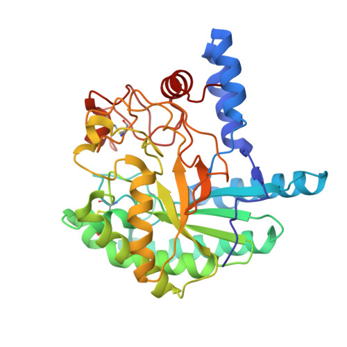
Structure of the Humicola Insolens Cellobiohydrolase Cel6A D416A Mutant in Complex with a Non-Hydrolysable Substrate Analogue, Methyl Cellobiosyl-4-Thio-Beta-Cellobioside, at 1.9 A

Varrot, A.Frandsen, T.Driguez, H.Davies, G.J.

(2002) Acta Crystallogr D Biol Crystallogr 58: 2201

  • DOI: https://doi.org/10.1107/s0907444902017006
  • Primary Citation of Related Structures:  
    1GZ1

  • PubMed Abstract: 

    The enzymatic degradation of cellulose continues to be one of the most important enzyme-catalysed reactions. Glycoside hydrolases from family GH-6 hydrolyse cellulose with inversion of the configuration of the anomeric carbon. Whilst the catalytic proton donor has been clearly identified (Asp226 in Humicola insolens Cel6A), the identification and even the existence of a potential Brønsted base remains unclear. Equally controversial is the role of surface-loop flexibility. Here, the structure of the D416A mutant of the H. insolens cellobiohydrolase Cel6A in complex with a non-hydrolysable thiooligosaccharide methyl cellobiosyl-4-thio-beta-cellobioside at 1.9 A resolution is presented. Substrate distortion in the -1 subsite, to a (2)S(0) skew-boat conformation, is observed, similar to that seen in the analogous Trichoderma reesei Cel6A structure [Zou et al. (1999), Structure, 7, 1035-1045], but the active-centre N-terminal loop of the H. insolens enzyme is found in a more open conformation than described for previous structures.


  • Organizational Affiliation

    Structural Biology Laboratory, Department of Chemistry, The University of York, Heslington, York Y010 5YW, England.


Macromolecules
Find similar proteins by:  (by identity cutoff)  |  3D Structure
Entity ID: 1
MoleculeChains Sequence LengthOrganismDetailsImage
CELLOBIOHYDROLASE II362Humicola insolensMutation(s): 0 
EC: 3.2.1.91
UniProt
Find proteins for Q9C1S9 (Humicola insolens)
Explore Q9C1S9 
Go to UniProtKB:  Q9C1S9
Entity Groups  
Sequence Clusters30% Identity50% Identity70% Identity90% Identity95% Identity100% Identity
UniProt GroupQ9C1S9
Sequence Annotations
Expand
  • Reference Sequence
Oligosaccharides

Help

Entity ID: 2
MoleculeChains Length2D Diagram Glycosylation3D Interactions
beta-D-glucopyranose-(1-4)-beta-D-glucopyranose-(1-4)-4-thio-beta-D-glucopyranose-(1-4)-methyl beta-D-glucopyranoside
B
4N/AN/A
Glycosylation Resources
GlyTouCan:  G15263TL
GlyCosmos:  G15263TL
Experimental Data & Validation

Experimental Data

  • Method: X-RAY DIFFRACTION
  • Resolution: 1.90 Å
  • R-Value Free: 0.211 
  • R-Value Work: 0.166 
  • R-Value Observed: 0.168 
  • Space Group: P 1 21 1
Unit Cell:
Length ( Å )Angle ( ˚ )
a = 48.257α = 90
b = 68.38β = 112.39
c = 55.18γ = 90
Software Package:
Software NamePurpose
REFMACrefinement
DENZOdata reduction
SCALEPACKdata scaling
AMoREphasing

Structure Validation

View Full Validation Report


Ligand Structure Quality Assessment 


Entry History 

Deposition Data

Revision History  (Full details and data files)

  • Version 1.0: 2003-02-12
    Type: Initial release
  • Version 1.1: 2011-10-19
    Changes: Derived calculations, Non-polymer description, Other, Structure summary, Version format compliance
  • Version 2.0: 2020-07-29
    Type: Remediation
    Reason: Carbohydrate remediation
    Changes: Atomic model, Data collection, Derived calculations, Other, Structure summary