1GA4

CRYSTAL STRUCTURE ANALYSIS OF PSCP (PSEUDOMONAS SERINE-CARBOXYL PROTEINASE) COMPLEXED WITH INHIBITOR PSEUDOIODOTYROSTATIN (THIS ENZYME RENAMED "SEDOLISIN" IN 2003)


Experimental Data Snapshot

  • Method: X-RAY DIFFRACTION
  • Resolution: 1.40 Å
  • R-Value Free: 0.155 
  • R-Value Work: 0.118 
  • R-Value Observed: 0.103 

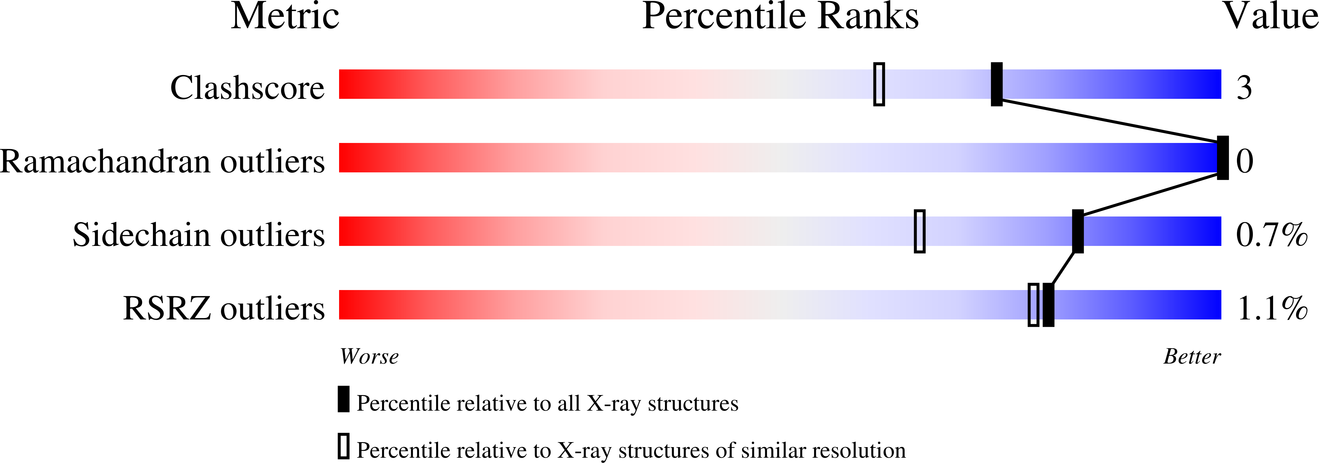
wwPDB Validation   3D Report Full Report


This is version 1.5 of the entry. See complete history


Literature

Carboxyl proteinase from Pseudomonas defines a novel family of subtilisin-like enzymes.

Wlodawer, A.Li, M.Dauter, Z.Gustchina, A.Uchida, K.Oyama, H.Dunn, B.M.Oda, K.

(2001) Nat Struct Biol 8: 442-446

  • DOI: https://doi.org/10.1038/87610
  • Primary Citation of Related Structures:  
    1GA4, 1GA6

  • PubMed Abstract: 

    The crystal structure of a pepstatin-insensitive carboxyl proteinase from Pseudomonas sp. 101 (PSCP) has been solved by single-wavelength anomalous diffraction using the absorption peak of bromide anions. Structures of the uninhibited enzyme and of complexes with an inhibitor that was either covalently or noncovalently bound were refined at 1.0-1.4 A resolution. The structure of PSCP comprises a single compact domain with a diameter of approximately 55 A, consisting of a seven-stranded parallel beta-sheet flanked on both sides by a number of helices. The fold of PSCP is a superset of the subtilisin fold, and the covalently bound inhibitor is linked to the enzyme through a serine residue. Thus, the structure of PSCP defines a novel family of serine-carboxyl proteinases (defined as MEROPS S53) with a unique catalytic triad consisting of Glu 80, Asp 84 and Ser 287.


  • Organizational Affiliation

    Protein Structure Section, Macromolecular Crystallography Laboratory, National Cancer Institute at Frederick, Maryland 21702, USA. wlodawer@ncifcrf.gov


Macromolecules
Find similar proteins by:  (by identity cutoff)  |  3D Structure
Entity ID: 1
MoleculeChains Sequence LengthOrganismDetailsImage
SERINE-CARBOXYL PROTEINASE372Pseudomonas sp.Mutation(s): 0 
EC: 3.4.23.37
UniProt
Find proteins for P42790 (Pseudomonas sp. (strain 101))
Explore P42790 
Go to UniProtKB:  P42790
Entity Groups  
Sequence Clusters30% Identity50% Identity70% Identity90% Identity95% Identity100% Identity
UniProt GroupP42790
Sequence Annotations
Expand
  • Reference Sequence

Find similar proteins by:  Sequence   |   3D Structure  

Entity ID: 2
MoleculeChains Sequence LengthOrganismDetailsImage
PSEUDOIODOTYROSTATINB [auth I]3N/AMutation(s): 0 
Sequence Annotations
Expand
  • Reference Sequence
Small Molecules
Modified Residues  2 Unique
IDChains TypeFormula2D DiagramParent
PHI
Query on PHI
B [auth I]L-PEPTIDE LINKINGC9 H10 I N O2PHE
TYB
Query on TYB
B [auth I]L-PEPTIDE LINKINGC9 H11 N O2TYR
Biologically Interesting Molecules (External Reference) 1 Unique
Experimental Data & Validation

Experimental Data

  • Method: X-RAY DIFFRACTION
  • Resolution: 1.40 Å
  • R-Value Free: 0.155 
  • R-Value Work: 0.118 
  • R-Value Observed: 0.103 
  • Space Group: P 62
Unit Cell:
Length ( Å )Angle ( ˚ )
a = 97.63α = 90
b = 97.63β = 90
c = 83.3γ = 120
Software Package:
Software NamePurpose
HKL-2000data collection
HKL-2000data reduction
SHELXL-97refinement
HKL-2000data scaling

Structure Validation

View Full Validation Report


Entry History 

Deposition Data

Revision History  (Full details and data files)

  • Version 1.0: 2000-12-13
    Type: Initial release
  • Version 1.1: 2008-04-27
    Changes: Version format compliance
  • Version 1.2: 2011-07-13
    Changes: Atomic model, Database references, Derived calculations, Non-polymer description, Structure summary, Version format compliance
  • Version 1.3: 2012-12-12
    Changes: Other
  • Version 1.4: 2017-10-04
    Changes: Advisory, Refinement description
  • Version 1.5: 2023-08-09
    Changes: Advisory, Data collection, Database references, Derived calculations, Refinement description