1G5G

FRAGMENT OF FUSION PROTEIN FROM NEWCASTLE DISEASE VIRUS


Experimental Data Snapshot

  • Method: X-RAY DIFFRACTION
  • Resolution: 3.30 Å
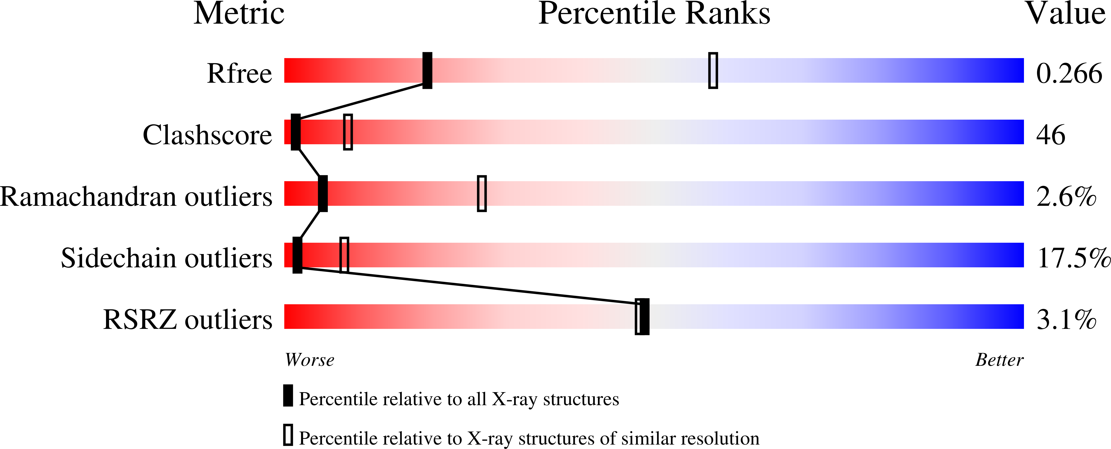
  • R-Value Free: 0.273 
  • R-Value Work: 0.224 

wwPDB Validation   3D Report Full Report


Ligand Structure Quality Assessment 


This is version 2.1 of the entry. See complete history


Literature

The structure of the fusion glycoprotein of Newcastle disease virus suggests a novel paradigm for the molecular mechanism of membrane fusion.

Chen, L.Gorman, J.J.McKimm-Breschkin, J.Lawrence, L.J.Tulloch, P.A.Smith, B.J.Colman, P.M.Lawrence, M.C.

(2001) Structure 9: 255-266

  • DOI: https://doi.org/10.1016/s0969-2126(01)00581-0
  • Primary Citation of Related Structures:  
    1G5G

  • PubMed Abstract: 

    Membrane fusion within the Paramyxoviridae family of viruses is mediated by a surface glycoprotein termed the "F", or fusion, protein. Membrane fusion is assumed to involve a series of structural transitions of F from a metastable (prefusion) state to a highly stable (postfusion) state. No detail is available at the atomic level regarding the metastable form of these proteins or regarding the transitions accompanying fusion.


  • Organizational Affiliation

    Biomolecular Research Institute, Parkville, Victoria 3052, Australia.


Macromolecules
Find similar proteins by:  (by identity cutoff)  |  3D Structure
Entity ID: 1
MoleculeChains Sequence LengthOrganismDetailsImage
Fusion glycoprotein F0
A, B, C, D, E
A, B, C, D, E, F
481avian paramyxovirus 1Mutation(s): 0 
UniProt
Find proteins for P35936 (Newcastle disease virus (strain D26/76))
Explore P35936 
Go to UniProtKB:  P35936
Entity Groups  
Sequence Clusters30% Identity50% Identity70% Identity90% Identity95% Identity100% Identity
UniProt GroupP35936
Sequence Annotations
Expand
  • Reference Sequence
Oligosaccharides

Help

Entity ID: 2
MoleculeChains Length2D Diagram Glycosylation3D Interactions
2-acetamido-2-deoxy-beta-D-glucopyranose-(1-4)-2-acetamido-2-deoxy-beta-D-glucopyranose
G, I, J, K, L
G, I, J, K, L, M, N
2N-Glycosylation
Glycosylation Resources
GlyTouCan:  G42666HT
GlyCosmos:  G42666HT
GlyGen:  G42666HT
Entity ID: 3
MoleculeChains Length2D Diagram Glycosylation3D Interactions
beta-D-mannopyranose-(1-4)-2-acetamido-2-deoxy-beta-D-glucopyranose-(1-4)-2-acetamido-2-deoxy-beta-D-glucopyranose
H
3N-Glycosylation
Glycosylation Resources
GlyTouCan:  G15407YE
GlyCosmos:  G15407YE
GlyGen:  G15407YE
Experimental Data & Validation

Experimental Data

  • Method: X-RAY DIFFRACTION
  • Resolution: 3.30 Å
  • R-Value Free: 0.273 
  • R-Value Work: 0.224 
  • Space Group: C 2 2 21
Unit Cell:
Length ( Å )Angle ( ˚ )
a = 134.39α = 90
b = 308.33β = 90
c = 243γ = 90
Software Package:
Software NamePurpose
DENZOdata reduction
SCALEPACKdata scaling
X-PLORmodel building
X-PLORrefinement
X-PLORphasing

Structure Validation

View Full Validation Report


Ligand Structure Quality Assessment 


Entry History 

Deposition Data

Revision History  (Full details and data files)

  • Version 1.0: 2002-02-27
    Type: Initial release
  • Version 1.1: 2008-04-27
    Changes: Version format compliance
  • Version 1.2: 2011-07-13
    Changes: Non-polymer description, Version format compliance
  • Version 1.3: 2018-01-31
    Changes: Experimental preparation
  • Version 2.0: 2020-07-29
    Type: Remediation
    Reason: Carbohydrate remediation
    Changes: Atomic model, Data collection, Database references, Derived calculations, Structure summary
  • Version 2.1: 2022-12-21
    Changes: Database references, Source and taxonomy, Structure summary