1F9K

WINGED BEAN ACIDIC LECTIN COMPLEXED WITH METHYL-ALPHA-D-GALACTOSE


Experimental Data Snapshot

  • Method: X-RAY DIFFRACTION
  • Resolution: 3.00 Å
  • R-Value Free: 0.251 
  • R-Value Work: 0.195 
  • R-Value Observed: 0.195 

wwPDB Validation   3D Report Full Report


Ligand Structure Quality Assessment 


This is version 2.2 of the entry. See complete history


Literature

Carbohydrate specificity and salt-bridge mediated conformational change in acidic winged bean agglutinin.

Manoj, N.Srinivas, V.R.Surolia, A.Vijayan, M.Suguna, K.

(2000) J Mol Biol 302: 1129-1137

  • DOI: https://doi.org/10.1006/jmbi.2000.4111
  • Primary Citation of Related Structures:  
    1F9K, 1FAY

  • PubMed Abstract: 

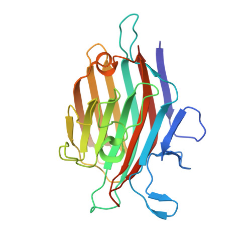
    Structures of two crystal forms of the dimeric acidic winged bean agglutinin (WBAII) complexed with methyl-alpha-D-galactose have been determined at 3.0 A and 3.3 A resolution. The subunit structure and dimerisation of the lectin are similar to those of the basic lectin from winged bean (WBAI) and the lectin from Erythrina corallodendron (EcorL). The conformation of a loop and its orientation with respect to the rest of the molecule in WBAII are, however, different from those in all the other legume lectins of known structure. This difference appears to have been caused by the formation of two strategically placed salt bridges in the former. Modelling based on the crystal structures provides a rationale for the specificity of the lectin, which is very different from that of WBAI, for the H-antigenic determinant responsible for O blood group reactivity. It also leads to a qualitative explanation for the thermodynamic data on sugar-binding to the lectin, with special emphasis on the role of a tyrosyl residue in the variable loop in the sugar-binding region in generating the carbohydrate specificity of WBAII.


  • Organizational Affiliation

    Molecular Biophysics Unit, Indian Institute of Science, Bangalore, India.


Macromolecules
Find similar proteins by:  (by identity cutoff)  |  3D Structure
Entity ID: 1
MoleculeChains Sequence LengthOrganismDetailsImage
ACIDIC LECTIN
A, B
238Psophocarpus tetragonolobusMutation(s): 3 
UniProt
Find proteins for Q9SM56 (Psophocarpus tetragonolobus)
Explore Q9SM56 
Go to UniProtKB:  Q9SM56
Entity Groups  
Sequence Clusters30% Identity50% Identity70% Identity90% Identity95% Identity100% Identity
UniProt GroupQ9SM56
Sequence Annotations
Expand
  • Reference Sequence
Oligosaccharides

Help

Entity ID: 2
MoleculeChains Length2D Diagram Glycosylation3D Interactions
alpha-D-mannopyranose-(1-4)-2-acetamido-2-deoxy-beta-D-glucopyranose-(1-4)-2-acetamido-2-deoxy-beta-D-glucopyranose
C, D
3N-Glycosylation
Glycosylation Resources
GlyTouCan:  G62182OO
GlyCosmos:  G62182OO
GlyGen:  G62182OO
Experimental Data & Validation

Experimental Data

  • Method: X-RAY DIFFRACTION
  • Resolution: 3.00 Å
  • R-Value Free: 0.251 
  • R-Value Work: 0.195 
  • R-Value Observed: 0.195 
  • Space Group: H 3
Unit Cell:
Length ( Å )Angle ( ˚ )
a = 182.11α = 90
b = 182.11β = 90
c = 44.99γ = 120
Software Package:
Software NamePurpose
AMoREphasing
CNSrefinement
DENZOdata reduction
SCALEPACKdata scaling

Structure Validation

View Full Validation Report


Ligand Structure Quality Assessment 


Entry History 

Deposition Data

Revision History  (Full details and data files)

  • Version 1.0: 2001-07-11
    Type: Initial release
  • Version 1.1: 2008-04-27
    Changes: Version format compliance
  • Version 1.2: 2011-07-13
    Changes: Non-polymer description, Version format compliance
  • Version 2.0: 2020-07-29
    Type: Remediation
    Reason: Carbohydrate remediation
    Changes: Advisory, Atomic model, Data collection, Database references, Derived calculations, Structure summary
  • Version 2.1: 2021-11-03
    Changes: Database references, Structure summary
  • Version 2.2: 2024-04-03
    Changes: Data collection, Refinement description