1EUO

Crystal structure of nitrophorin 2 (prolixin-S)


Experimental Data Snapshot

  • Method: X-RAY DIFFRACTION
  • Resolution: 2.00 Å
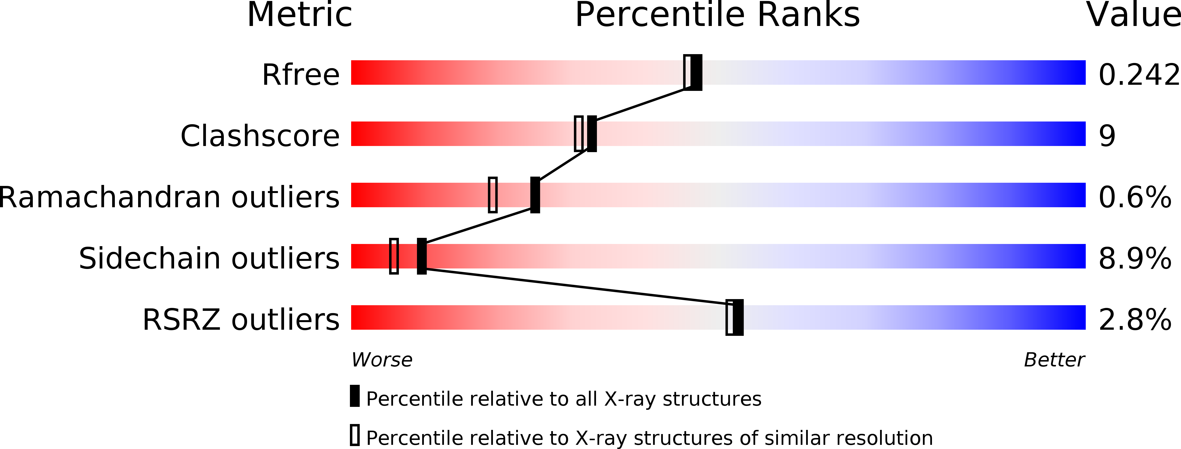
  • R-Value Free: 0.246 
  • R-Value Work: 0.195 
  • R-Value Observed: 0.197 

wwPDB Validation   3D Report Full Report


Ligand Structure Quality Assessment 


This is version 1.4 of the entry. See complete history


Literature

The crystal structure of nitrophorin 2. A trifunctional antihemostatic protein from the saliva of Rhodnius prolixus

Andersen, J.F.Montfort, W.R.

(2000) J Biol Chem 275: 30496-30503

  • DOI: https://doi.org/10.1074/jbc.M002857200
  • Primary Citation of Related Structures:  
    1EUO

  • PubMed Abstract: 

    Nitrophorin 2 (NP2) (also known as prolixin-S) is a salivary protein that transports nitric oxide, binds histamine, and acts as an anticoagulant during blood feeding by the insect Rhodnius prolixus. The 2.0-A crystal structure of NP2 reveals an eight-stranded antiparallel beta-barrel containing a ferric heme coordinated through His(57), similar to the structures of NP1 and NP4. All four Rhodnius nitrophorins transport NO and sequester histamine through heme binding, but only NP2 acts as an anticoagulant. Here, we demonstrate that recombinant NP2, but not recombinant NP1 or NP4, is a potent anticoagulant; recombinant NP3 also displays minor activity. Comparison of the nitrophorin structures suggests that a surface region near the C terminus and the loops between beta strands B-C and E-F is responsible for the anticoagulant activity. NP2 also displays larger NO association rates and smaller dissociation rates than NP1 and NP4, which may result from a more open and more hydrophobic distal pocket, allowing more rapid solvent reorganization on ligand binding. The NP2 protein core differs from NP1 and NP4 in that buried Glu(53), which allows for larger NO release rates when deprotonated, hydrogen bonds to invariant Tyr(81). Surprisingly, this tyrosine lies on the protein surface in NP1 and NP4.


  • Organizational Affiliation

    Department of Biochemistry, University of Arizona, Tucson, Arizona 85721, USA.


Macromolecules
Find similar proteins by:  (by identity cutoff)  |  3D Structure
Entity ID: 1
MoleculeChains Sequence LengthOrganismDetailsImage
NITROPHORIN 2180Rhodnius prolixusMutation(s): 0 
Gene Names: SALIVARY GLAND CDNA
UniProt
Find proteins for Q26241 (Rhodnius prolixus)
Explore Q26241 
Go to UniProtKB:  Q26241
Entity Groups  
Sequence Clusters30% Identity50% Identity70% Identity90% Identity95% Identity100% Identity
UniProt GroupQ26241
Sequence Annotations
Expand
  • Reference Sequence
Small Molecules
Ligands 2 Unique
IDChains Name / Formula / InChI Key2D Diagram3D Interactions
HEM
Query on HEM

Download Ideal Coordinates CCD File 
B [auth A]PROTOPORPHYRIN IX CONTAINING FE
C34 H32 Fe N4 O4
KABFMIBPWCXCRK-RGGAHWMASA-L
NH3
Query on NH3

Download Ideal Coordinates CCD File 
C [auth A]AMMONIA
H3 N
QGZKDVFQNNGYKY-UHFFFAOYSA-N
Experimental Data & Validation

Experimental Data

  • Method: X-RAY DIFFRACTION
  • Resolution: 2.00 Å
  • R-Value Free: 0.246 
  • R-Value Work: 0.195 
  • R-Value Observed: 0.197 
  • Space Group: P 21 21 2
Unit Cell:
Length ( Å )Angle ( ˚ )
a = 40.34α = 90
b = 127.96β = 90
c = 33.72γ = 90
Software Package:
Software NamePurpose
MADNESSdata collection
SCALAdata scaling
X-PLORmodel building
CNSrefinement
MADNESSdata reduction
CCP4data scaling
X-PLORphasing

Structure Validation

View Full Validation Report


Ligand Structure Quality Assessment 


Entry History 

Deposition Data

Revision History  (Full details and data files)

  • Version 1.0: 2000-05-10
    Type: Initial release
  • Version 1.1: 2008-04-27
    Changes: Version format compliance
  • Version 1.2: 2011-07-13
    Changes: Version format compliance
  • Version 1.3: 2017-10-04
    Changes: Refinement description
  • Version 1.4: 2018-04-18
    Changes: Data collection