1ETP

CRYSTAL STRUCTURE OF CYTOCHROME C4 FROM PSEUDOMONAS STUTZERI


Experimental Data Snapshot

  • Method: X-RAY DIFFRACTION
  • Resolution: 2.20 Å
  • R-Value Free: 0.267 
  • R-Value Work: 0.201 
  • R-Value Observed: 0.201 

wwPDB Validation   3D Report Full Report


Ligand Structure Quality Assessment 


This is version 1.2 of the entry. See complete history


Literature

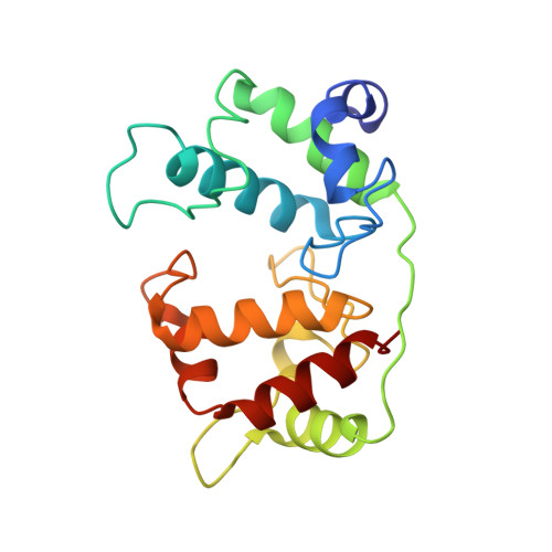
Crystal structure of the dihaem cytochrome c4 from Pseudomonas stutzeri determined at 2.2A resolution.

Kadziola, A.Larsen, S.

(1997) Structure 5: 203-216

  • DOI: https://doi.org/10.1016/s0969-2126(97)00179-2
  • Primary Citation of Related Structures:  
    1ETP

  • PubMed Abstract: 

    . Cytochromes c4 are dihaem cytochromes c found in a variety of bacteria. They are assumed to take part in the electron-transport systems associated with both aerobic and anaerobic respiration. The cytochrome c4 proteins are located in the periplasm, predominantly bound to the inner membrane, and are able to transfer electrons between membrane-bound reduction systems and terminal oxidases. Alignment of cytochrome c4 sequences from three bacteria, Pseudomonas aeruginosa, Pseudomonas stutzeri and Azotobacter vinelandii, suggests that these dihaem proteins are composed of two similar domains. Two distinctly different redox potentials have been measured for the Ps. stutzeri cytochrome c4, however.


  • Organizational Affiliation

    Centre for Crystallographic Studies, Department of Chemistry, University of Copenhagen, Universitetsparken 5, DK-2100 Copenhagen, Denmark.


Macromolecules
Find similar proteins by:  (by identity cutoff)  |  3D Structure
Entity ID: 1
MoleculeChains Sequence LengthOrganismDetailsImage
CYTOCHROME C4
A, B
190Stutzerimonas stutzeriMutation(s): 0 
UniProt
Find proteins for Q52369 (Stutzerimonas stutzeri)
Explore Q52369 
Go to UniProtKB:  Q52369
Entity Groups  
Sequence Clusters30% Identity50% Identity70% Identity90% Identity95% Identity100% Identity
UniProt GroupQ52369
Sequence Annotations
Expand
  • Reference Sequence
Experimental Data & Validation

Experimental Data

  • Method: X-RAY DIFFRACTION
  • Resolution: 2.20 Å
  • R-Value Free: 0.267 
  • R-Value Work: 0.201 
  • R-Value Observed: 0.201 
  • Space Group: P 1 21 1
Unit Cell:
Length ( Å )Angle ( ˚ )
a = 49.49α = 90
b = 58.58β = 96.96
c = 63.51γ = 90
Software Package:
Software NamePurpose
X-PLORmodel building
X-PLORrefinement
DENZOdata reduction
X-PLORphasing

Structure Validation

View Full Validation Report


Ligand Structure Quality Assessment 


Entry History 

Deposition Data

Revision History  (Full details and data files)

  • Version 1.0: 1997-02-12
    Type: Initial release
  • Version 1.1: 2008-03-24
    Changes: Version format compliance
  • Version 1.2: 2011-07-13
    Changes: Version format compliance