1ESG

RESTRICTION ENDONUCLEASE BAMHI BOUND TO A NON-SPECIFIC DNA.


Experimental Data Snapshot

  • Method: X-RAY DIFFRACTION
  • Resolution: 1.90 Å
  • R-Value Free: 0.230 
  • R-Value Work: 0.191 

wwPDB Validation   3D Report Full Report


This is version 1.4 of the entry. See complete history


Literature

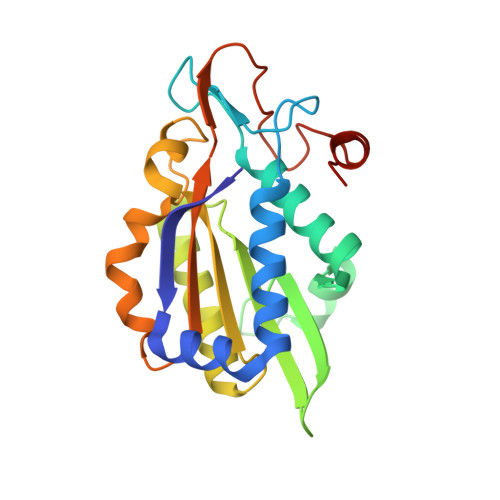
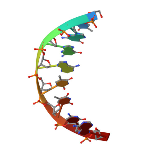
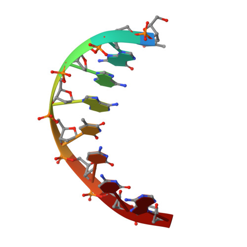
Structure of BamHI bound to nonspecific DNA: a model for DNA sliding.

Viadiu, H.Aggarwal, A.K.

(2000) Mol Cell 5: 889-895

  • DOI: https://doi.org/10.1016/s1097-2765(00)80329-9
  • Primary Citation of Related Structures:  
    1ESG

  • PubMed Abstract: 

    The central problem faced by DNA binding proteins is how to select the correct DNA sequence from the sea of nonspecific sequences in a cell. The problem is particularly acute for bacterial restriction enzymes because cleavage at an incorrect DNA site could be lethal. To understand the basis of this selectivity, we report here the crystal structure of endonuclease BamHI bound to noncognate DNA. We show that, despite only a single base pair change in the recognition sequence, the enzyme adopts an open configuration that is on the pathway between free and specifically bound forms of the enzyme. Surprisingly, the DNA drops out of the binding cleft with a total loss of base-specific and backbone contacts. Taken together, the structure provides a remarkable snapshot of an enzyme poised for linear diffusion (rather than cleavage) along the DNA.


  • Organizational Affiliation

    Department of Biochemistry and Molecular Biophysics, Columbia University, New York, New York 10032, USA.


Macromolecules

Find similar proteins by:  (by identity cutoff)  |  3D Structure
Entity ID: 3
MoleculeChains Sequence LengthOrganismDetailsImage
TYPE II RESTRICTION ENZYME BAMHIC [auth A],
D [auth B]
213Bacillus amyloliquefaciensMutation(s): 0 
EC: 3.1.21.4
UniProt
Find proteins for P23940 (Bacillus amyloliquefaciens)
Explore P23940 
Go to UniProtKB:  P23940
Entity Groups  
Sequence Clusters30% Identity50% Identity70% Identity90% Identity95% Identity100% Identity
UniProt GroupP23940
Sequence Annotations
Expand
  • Reference Sequence

Find similar nucleic acids by:  Sequence   |   3D Structure  

Entity ID: 1
MoleculeChains LengthOrganismImage
DNA (5'-D(*TP*GP*GP*AP*TP*TP*CP*A)-3')A [auth C]8N/A
Sequence Annotations
Expand
  • Reference Sequence

Find similar nucleic acids by:  Sequence   |   3D Structure  

Entity ID: 2
MoleculeChains LengthOrganismImage
DNA (5'-D(*TP*GP*AP*AP*TP*CP*CP*A)-3')B [auth D]8N/A
Sequence Annotations
Expand
  • Reference Sequence
Experimental Data & Validation

Experimental Data

  • Method: X-RAY DIFFRACTION
  • Resolution: 1.90 Å
  • R-Value Free: 0.230 
  • R-Value Work: 0.191 
  • Space Group: P 21 21 21
Unit Cell:
Length ( Å )Angle ( ˚ )
a = 114.8α = 90
b = 91.1β = 90
c = 66.4γ = 90
Software Package:
Software NamePurpose
AMoREphasing
CNSrefinement
DENZOdata reduction
SCALEPACKdata scaling

Structure Validation

View Full Validation Report


Entry History 

Deposition Data

Revision History  (Full details and data files)

  • Version 1.0: 2000-05-31
    Type: Initial release
  • Version 1.1: 2008-04-27
    Changes: Version format compliance
  • Version 1.2: 2011-07-13
    Changes: Version format compliance
  • Version 1.3: 2018-01-31
    Changes: Experimental preparation
  • Version 1.4: 2024-02-07
    Changes: Data collection, Database references