1DUW

STRUCTURE OF NONAHEME CYTOCHROME C


Experimental Data Snapshot

  • Method: X-RAY DIFFRACTION
  • Resolution: 1.89 Å
  • R-Value Free: 0.235 
  • R-Value Work: 0.183 
  • R-Value Observed: 0.183 

wwPDB Validation   3D Report Full Report


Ligand Structure Quality Assessment 


This is version 2.0 of the entry. See complete history


Literature

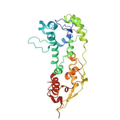
Three-dimensional structure of the nonaheme cytochrome c from Desulfovibrio desulfuricans Essex in the Fe(III) state at 1.89 A resolution.

Umhau, S.Fritz, G.Diederichs, K.Breed, J.Welte, W.Kroneck, P.M.

(2001) Biochemistry 40: 1308-1316

  • DOI: https://doi.org/10.1021/bi001479a
  • Primary Citation of Related Structures:  
    1DUW

  • PubMed Abstract: 

    A nine heme group containing cytochrome c isolated from the soluble and membrane fractions of Desulfovibrio desulfuricans Essex, termed nonaheme cytochrome c, was crystallized, and the structure was solved using the multiple wavelength anomalous dispersion (MAD) phasing method. Refinement was carried out to a resolution of 1.89 A, and anisotropic temperature factors were addressed to the iron and sulfur atoms in the model. The structure revealed two cytochrome c(3) like domains with the typical arrangement of four heme centers. Both domains flanked an extra heme buried under the protein surface. This heme is held in position by loop extensions in each of the two domains. Although both the N- and C-terminal tetraheme domains exhibit a fold and heme arrangement very similar to that of cytochrome c(3), they differ considerably in their loop extensions and electrostatic surface. Analysis of the structure provides evidence for a different function of both domains, namely, anchoring the protein in a transmembranous complex with the N-terminal domain and formation of an electron-transfer complex with hydrogenase by the C-terminal domain.


  • Organizational Affiliation

    Fachbereich Biologie, Mathematisch-Naturwissenschaftliche Sektion, Universität Konstanz, 78457 Konstanz, Germany.


Macromolecules
Find similar proteins by:  (by identity cutoff)  |  3D Structure
Entity ID: 1
MoleculeChains Sequence LengthOrganismDetailsImage
NONAHEME CYTOCHROME C292Desulfovibrio desulfuricansMutation(s): 0 
Membrane Entity: Yes 
UniProt
Find proteins for Q9XCU0 (Desulfovibrio desulfuricans)
Explore Q9XCU0 
Go to UniProtKB:  Q9XCU0
Entity Groups  
Sequence Clusters30% Identity50% Identity70% Identity90% Identity95% Identity100% Identity
UniProt GroupQ9XCU0
Sequence Annotations
Expand
  • Reference Sequence
Small Molecules
Ligands 2 Unique
IDChains Name / Formula / InChI Key2D Diagram3D Interactions
HEC
Query on HEC

Download Ideal Coordinates CCD File 
B [auth A]
C [auth A]
D [auth A]
E [auth A]
F [auth A]
B [auth A],
C [auth A],
D [auth A],
E [auth A],
F [auth A],
G [auth A],
H [auth A],
I [auth A],
J [auth A]
HEME C
C34 H34 Fe N4 O4
HXQIYSLZKNYNMH-LJNAALQVSA-N
GOL
Query on GOL

Download Ideal Coordinates CCD File 
K [auth A]GLYCEROL
C3 H8 O3
PEDCQBHIVMGVHV-UHFFFAOYSA-N
Experimental Data & Validation

Experimental Data

  • Method: X-RAY DIFFRACTION
  • Resolution: 1.89 Å
  • R-Value Free: 0.235 
  • R-Value Work: 0.183 
  • R-Value Observed: 0.183 
  • Space Group: P 41 21 2
Unit Cell:
Length ( Å )Angle ( ˚ )
a = 55.4α = 90
b = 55.4β = 90
c = 236.8γ = 90
Software Package:
Software NamePurpose
SOLVEphasing
SHELXL-97refinement
MAR345data collection
XDSdata scaling

Structure Validation

View Full Validation Report


Ligand Structure Quality Assessment 


Entry History 

Deposition Data

Revision History  (Full details and data files)

  • Version 1.0: 2001-03-21
    Type: Initial release
  • Version 1.1: 2008-04-27
    Changes: Version format compliance
  • Version 1.2: 2011-07-13
    Changes: Derived calculations, Version format compliance
  • Version 1.3: 2017-10-04
    Changes: Refinement description
  • Version 1.4: 2018-01-31
    Changes: Experimental preparation
  • Version 2.0: 2021-03-03
    Changes: Advisory, Atomic model, Data collection, Derived calculations, Non-polymer description, Structure summary